Reasons for choosing our testing services
ZHONGXI Testing has obtained inspection qualification certifications from multiple countries and regions worldwide. We possess a senior testing team and advanced testing methods, providing independent, impartial, and professional third-party verification services for global carbon projects.

Internationally recognized authority
Certified by multiple international standards such as CNAS, VCS, and GS, with reports universally applicable worldwide.

Global service capability
Covering 140+ countries and regions, it supports on-site detection and remote verification in multiple languages.

Professional experimental methods
Adopt standard experimental methods to ensure accurate and reliable data.
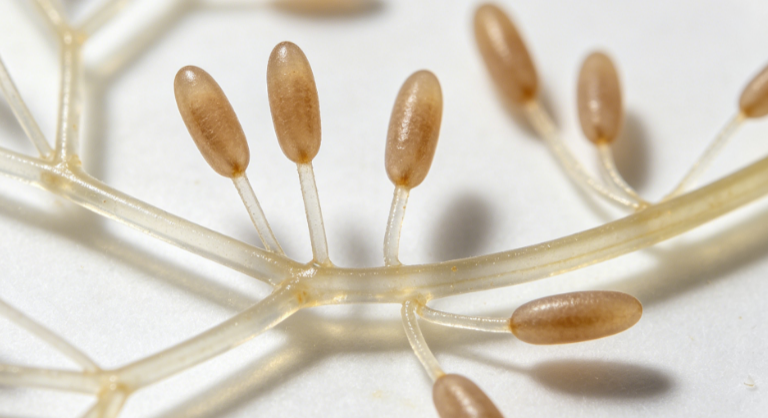
Professional Morphological Characterization Service for Nothophoma (or Pseudocercospora) from Alligator Weed – Accurate Fungal Identification
We understand that you are searching for morphological characterisation of the fungus associated with Alternanthera philoxeroides (alligator weed) – commonly referred to as “莲子草假格孢菌” – because you need to confirm species identity, distinguish it from look‑alike pathogens, document strain features for biocontrol development, or meet taxonomic reporting requirements for research or regulatory submission. Our comprehensive, high‑resolution morphological analysis platform goes far beyond basic light microscopy. We deliver detailed characterisation of conidial, hyphal, and sporodochial/pycnidial structures, including ultrastructural features, morphometric statistics, and cultural characteristics, enabling definitive identification and comparison with type descriptions.
What We Examine – From Colony Morphology to Ultrastructural Details
Our standard characterisation includes: colony appearance on multiple media (PDA, MEA, OA, and host leaf extract agar) – colour, texture, margin, zonation, and reverse pigmentation. Using brightfield, phase‑contrast, and differential interference contrast (DIC) microscopy, we capture hyphal diameter, septation, branching patterns, and presence of chlamydospores. For sporulating structures, we measure: conidiophores (length, width, branching, geniculation, scars), conidiogenous cells (type: phialidic, annellidic, or sympodial), and conidia (shape, size range, septation number, wall ornamentation, hila, and germination pores). We also examine pycnidia or acervuli (if applicable) – diameter, ostiole, wall layers, and setae. Each measurement is based on at least 50 randomly selected structures from multiple replicates, with statistical reporting (mean ± SD, min‑max, 95% CI).
Advanced Capabilities for Definitive Identification
We go beyond routine light microscopy. Our scanning electron microscopy (SEM) at magnifications up to 50,000× reveals surface ornamentation of conidia and conidiophores (verrucose, echinulate, smooth, or striate) – critical for distinguishing Nothophoma, Pseudocercospora, Alternaria-like taxa, and other coelomycetes or hyphomycetes. We also offer transmission electron microscopy (TEM) for septal pore ultrastructure and cell wall layering when required. For living cultures, we perform time‑lapse microscopy to document sporulation ontogeny (e.g., blastic vs. thallic conidial development). In addition, we provide slide culture preparation to observe undisturbed sporulation patterns. All images are captured with calibrated digital microscopy and delivered with scale bars, annotation, and high‑resolution TIFF files.
What You Receive – A Complete Morphological Report
Your final report includes: a full written description following mycological standards (e.g., format of Studies in Mycology or Persoonia), detailed measurement tables for all structures, comparative tables against closely related taxa (based on published type descriptions), and a decision on tentative identification. We also provide a photographic plate with labelled figures: colony obverse/reverse, microscopic structures at multiple magnifications (DIC, phase, SEM), and any distinctive features (e.g., conidial chains, beak formation, setae). For strains intended for biocontrol or patent deposit, we include a diagnostic morphological key differentiating your isolate from known saprophytes or pathogens of the same host.
Our Distinct Advantages in Morphological Characterisation
1. Specialist expertise in “hyphomycete and coelomycete” fungi: Our team includes mycologists who have described new species from subtropical weeds and are familiar with the subtle morphological differences between Pseudocercospora, Cercospora, Nothophoma, Phoma‑like, and Alternaria‑sectioned genera. 2. Multi‑method validation: We cross‑correlate light microscopy, SEM, and cultural data to avoid artefacts from mounting media or culture age. 3. High‑throughput slide scanning: Our automated slide scanner captures entire mounts at 40× and 100× oil, allowing you to review the whole preparation remotely. 4. Preservation of voucher specimens: We prepare and deposit permanent slides (in PVLG, lactofuchsin, or glycerol‑jelly) and dried herbarium specimens of your culture on host leaf or agar block – ready for deposition in a recognised herbarium (e.g., BPI, IMI, HMAS). 5. Integration with molecular data (optional): While this service is morphological, we can combine it with ITS, LSU, tef1, or tub2 sequencing (additional fee) to provide a polyphasic identification – highly recommended for publication or regulatory submission.
Why Choose Our Service Over General Microbiology Labs
Most routine labs stop at “hyaline septate hyphae” or “conidia observed” without measuring or imaging. We give you taxonomically robust characterisation that can stand alone or complement molecular data. Our reports have been used for fungal identification for biocontrol agent registration, distinguishing new species, and quality control of commercial fungal products. We provide one‑on‑one consultation to interpret ambiguous structures and advise on optimal culture conditions (light, temperature, media) to induce sporulation – because many “假格孢菌” are slow to sporulate on standard media. No hidden fees for SEM or advanced stainings; we deliver a transparent, defensible report.
Ready to Characterise Your Alligator Weed Fungus Morphologically?
Whether you need to confirm the identity of a potential biocontrol agent, document a new host record, or resolve a taxonomic dispute, our morphological service provides the precision and depth you require. Contact us today to discuss your isolate – we accept living pure cultures, dried herbarium fragments, or fresh infected leaf samples. We offer a free preliminary consultation, guidelines for subculturing and shipment, and a discounted pilot analysis for first‑time clients. Let us help you see the structures that define your fungus.