Reasons for choosing our testing services
ZHONGXI Testing has obtained inspection qualification certifications from multiple countries and regions worldwide. We possess a senior testing team and advanced testing methods, providing independent, impartial, and professional third-party verification services for global carbon projects.

Internationally recognized authority
Certified by multiple international standards such as CNAS, VCS, and GS, with reports universally applicable worldwide.

Global service capability
Covering 140+ countries and regions, it supports on-site detection and remote verification in multiple languages.

Professional experimental methods
Adopt standard experimental methods to ensure accurate and reliable data.
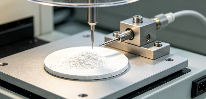
Comprehensive Physicochemical and Morphological Characterisation of Nano‑Magnesium Oxide: A Specialized Analytical Service for Advanced Functional Materials
Nano‑magnesium oxide (nano‑MgO) has emerged as a versatile material in catalysis, antimicrobial coatings, advanced ceramics, and flame‑retardant polymer composites, owing to its high surface area, unique surface reactivity, and wide bandgap. However, the functional performance of nano‑MgO is exquisitely sensitive to a range of nanoscale parameters, including primary crystallite size, agglomerate distribution, specific surface area, surface hydroxyl density, trace elemental impurities, and crystalline perfection. Clients seeking testing for nano‑MgO typically face challenges related to batch‑to‑batch variability in catalytic activity, inconsistent dispersion in polymer matrices, unanticipated cytotoxicity from impurity leaching, or phase instability during high‑temperature processing. Our laboratory has established a fully integrated, multi‑technique analytical platform that combines high‑resolution electron microscopy, surface area analysis, advanced diffraction, and ultra‑trace spectroscopic profiling, delivering a definitive, process‑relevant fingerprint that enables manufacturers and researchers to control synthesis, ensure reproducibility, and qualify nano‑MgO for the most demanding applications.
Precision Particle Size, Morphology, and Dispersion State
The primary particle size and agglomeration state of nano‑MgO directly influence its reactivity, optical properties, and reinforcing efficiency. We employ a combination of transmission electron microscopy (TEM) at 300 kV with aberration correction and high‑resolution scanning electron microscopy (FE‑SEM) to obtain direct measurements of primary crystallite diameter, shape distribution, and lattice fringe imaging with a spatial resolution of 0.08 nm. Using automated image analysis of >1,000 particles per sample, we report the mean diameter, standard deviation, and diameter distribution (D10, D50, D90) with repeatability of < 0.2 nm. For the agglomerate size in suspension, we use dynamic light scattering (DLS) and nanoparticle tracking analysis (NTA) in various dispersion media (water, ethanol, NMP) to provide hydrodynamic size distribution and polydispersity index (PdI) with precision < 1% RSD. We also assess the dispersibility and sedimentation stability via multiple‑sample light scattering (Turbiscan), providing a stability index that predicts formulation behaviour. These complementary techniques ensure that both the primary nanocrystal and its agglomerate state are fully characterised.
High‑Precision Specific Surface Area and Porosity
The high specific surface area (SSA) of nano‑MgO is the primary driver of its adsorption and catalytic properties. We measure BET specific surface area by nitrogen physisorption at 77 K using a multi‑point method over a relative pressure range of 0.01–0.30, achieving a reproducibility of < 0.5% RSD and a detection limit of 0.01 m²/g. For comprehensive pore architecture, we also acquire full adsorption‑desorption isotherms up to P/P₀ = 0.995 and apply DFT/NLDFT models with slit‑cylindrical pore geometries to obtain the micropore volume (t‑plot), mesopore size distribution (BJH), and total pore volume from 0.4 nm to 50 nm. For macroporosity, we use mercury intrusion porosimetry (MIP) up to 60,000 psi to determine the intra‑aggregate porosity and inter‑particle void volume. Our SSA and porosity data are cross‑validated with helium pycnometry for skeletal density, ensuring consistency across the textural characterisation.
Crystalline Phase Purity, Crystallite Size, and Lattice Strain
The cubic (periclase) phase of MgO is the desired structure, but the presence of hydrates (brucite, Mg(OH)₂) or carbonates (MgCO₃) can severely alter surface chemistry. We use high‑resolution powder X‑ray diffraction (HR‑XRD) with Cu Kα radiation and a step size of 0.003° 2θ, applying Rietveld refinement to quantify the relative fractions of periclase, brucite, and any other crystalline impurities with an accuracy of ±0.3 wt% and a detection limit of < 0.5 wt%. We also determine the crystallite size (via Scherrer and Williamson‑Hall methods) and the microstrain, which are directly correlated with the surface defect density and catalytic activity. For amorphous surface layers or disordered regions, we complement the XRD with Raman microspectroscopy (532 nm and 785 nm excitation) and solid‑state 25Mg magic‑angle spinning NMR to probe the local coordination environment and to identify any non‑periclase magnesium sites. This comprehensive phase and microstructure profile ensures that your nano‑MgO is both chemically and structurally well‑defined.
Surface Chemistry and Hydroxyl Group Density
The surface of nano‑MgO is typically terminated with hydroxyl groups, which mediate its interaction with adsorbates, polymers, and biological interfaces. We quantify the surface hydroxyl density using a combination of Thermogravimetric Analysis (TGA) under nitrogen from 30 °C to 800 °C (mass loss corresponding to dehydroxylation) and temperature‑programmed desorption (TPD) of water coupled with mass spectrometry, achieving a detection limit of 0.05 mmol OH/g and a repeatability of < 2% RSD. For chemical speciation, we use X‑ray photoelectron spectroscopy (XPS) with depth profiling to obtain the O 1s core‑level spectra, deconvoluting the contributions from lattice oxygen (O²⁻), hydroxyl (‑OH), and adsorbed water (H₂O). We also characterise the surface acidity/basicity via potentiometric titration and pyridine adsorption FTIR (Py‑FTIR), providing the number and strength of Lewis acidic and basic sites. This surface profile is essential for predicting the material's performance in catalysis, adsorption, and composite applications.
Ultra‑Trace Elemental Purity and Leachable Impurity Profiling
Even at sub‑ppm levels, transition metals (Fe, Cu, Ni, Mn, Cr) and alkali metals (Na, K) can significantly alter the electrical, catalytic, and toxicological properties of nano‑MgO. We digest the material in microwave‑assisted high‑purity acids and analyse by inductively coupled plasma tandem mass spectrometry (ICP‑MS/MS) with collision/reaction cell (O₂, NH₃, H₂) to eliminate polyatomic interferences (e.g., 40Ar16O⁺ on 56Fe, 40Ar35Cl⁺ on 75As) and achieve detection limits of 0.01–0.5 ppb for over 50 elements. For halogens (Cl, F, Br) and sulfur, we use ion chromatography (IC) after alkaline extraction and combustion‑infrared detection, respectively. We also perform leaching tests in simulated biological or environmental media (e.g., phosphate‑buffered saline, acidic water) to quantify the bioaccessible or environmental release fraction of metal ions, which is critical for biomedical and environmental applications. Our impurity report includes expanded uncertainties (k=2) and pass/fail status against the most stringent semiconductor‑grade, pharmaceutical‑grade, or industrial specifications.
Thermal Stability and Phase Transformation Behaviour
Nano‑MgO can undergo sintering, grain growth, and phase transformation (to Mg(OH)₂ or even MgO·H₂O) under humid or high‑temperature conditions. We perform simultaneous thermogravimetric and differential scanning calorimetry (TGA‑DSC) from 30 °C to 1000 °C under air, nitrogen, and steam‑containing atmospheres at heating rates of 2, 5, and 10 °C/min. We determine the temperature of dehydration (loss of adsorbed water), dehydroxylation (loss of structural OH), and any carbonation/decarbonation steps. For in situ structural evolution, we use high‑temperature XRD (HT‑XRD) up to 800 °C to monitor crystallite growth, lattice parameter changes, and the onset of sintering. We also conduct isothermal ageing at 200 °C, 400 °C, and 600 °C for up to 24 hours, followed by BET and TEM re‑characterisation, to provide a thermal stability limit for your specific application. The combined thermal profile enables you to define safe processing and service temperature windows.
Dispersion and Zeta Potential for Colloidal Stability
For liquid‑based formulations, the colloidal stability of nano‑MgO is governed by its zeta potential and surface charge. We measure zeta potential by electrophoretic light scattering (ELS) over a pH range of 2–12 and at varying ionic strengths (0.001–0.1 M), using automatic titrations with a precision of ±0.5 mV. From the zeta potential vs. pH curve, we determine the isoelectric point (IEP), which is critical for predicting aggregation and deposition behaviour. We also perform dynamic light scattering (DLS) at multiple concentrations to evaluate the critical agglomeration concentration (CAC) and to assess the effectiveness of common dispersants (e.g., surfactants, polymers). This colloidal fingerprint is indispensable for formulating stable suspensions for coatings, catalyst slurries, or nanocomposites.
Accelerated Stability and Shelf‑Life Testing
Nano‑MgO is sensitive to atmospheric moisture and CO₂, which can convert its surface to magnesium hydroxide and carbonate, reducing its activity. We conduct accelerated ageing tests at 40 °C/75% RH, 60 °C/ambient, and under CO₂‑enriched atmosphere (5000 ppm) for up to 6 months, with periodic re‑analysis of BET area, hydroxyl group density, XRD phase composition, and zeta potential. The degradation kinetics are modelled using Avrami‑Erofeev and diffusion‑controlled equations to estimate the shelf‑life under recommended storage conditions (dry, inert, sealed). We also evaluate the effect of different packaging materials (e.g., multi‑layer foil bags vs. HDPE containers) and provide specific recommendations to preserve the nanomaterial's properties.
Our Distinctive Competencies and Analytical Superiority
Our service is uniquely distinguished by the orthogonal, fully traceable integration of HR‑TEM/STEM, BET with DFT, HR‑XRD with Rietveld refinement, XPS surface chemical profiling, ICP‑MS/MS ultra‑trace impurity analysis, TGA‑DSC‑EGA‑MS thermal characterisation, and colloidal stability assessment—all performed on the same representative sample to eliminate cross‑batch variability and to enable direct, multivariate correlations (e.g., crystallite size vs. surface area, impurity content vs. thermal stability, or zeta potential vs. dispersion stability). We operate under ISO/IEC 17025 accreditation and maintain in‑house reference nano‑MgO materials (with certified particle size and surface area) that are periodically cross‑checked with NIST SRM 2600 (magnesia) and other international standards. Our proprietary “Nano‑MgO Quality and Performance Index” (NMPI™) combines primary particle size, BET area, phase purity, impurity sum, and dehydroxylation temperature into a single numerical score that predicts catalytic activity, thermal stability, and dispersion behaviour. This index has been validated against >30 commercial and research‑grade nano‑MgO samples.
We achieve exceptional precision: < 0.2 nm for TEM diameter, < 0.5% RSD for BET area, < 0.3 wt% for phase fraction, < 0.5 ppb for critical metals, and < 0.5 mV for zeta potential. Our turnaround time for the full characterisation suite (including accelerated stability tests) is 10–14 working days, with expedited 5‑day service for urgent material qualification. Crucially, our team of PhD‑level nanomaterial scientists, surface chemists, and catalytic engineers provides a comprehensive interpretative report that translates each parameter into actionable guidance—e.g., how to control calcination temperature to avoid crystallite growth, how to identify the root cause of batch‑to‑batch surface area variations, or how to select the appropriate surface treatment to enhance dispersibility. With over 20 successful projects on nano‑MgO and related alkaline earth oxide nanomaterials, we empower our clients to achieve synthesis reproducibility, optimise functional performance, and confidently scale from laboratory to production—all with the highest level of scientific rigour and technical credibility.