Reasons for choosing our testing services
ZHONGXI Testing has obtained inspection qualification certifications from multiple countries and regions worldwide. We possess a senior testing team and advanced testing methods, providing independent, impartial, and professional third-party verification services for global carbon projects.

Internationally recognized authority
Certified by multiple international standards such as CNAS, VCS, and GS, with reports universally applicable worldwide.

Global service capability
Covering 140+ countries and regions, it supports on-site detection and remote verification in multiple languages.

Professional experimental methods
Adopt standard experimental methods to ensure accurate and reliable data.
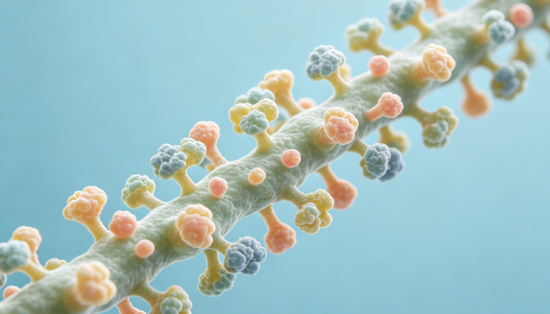
Neural Cell Adhesion Molecule (NCAM) Testing – Quantification, Isoform Profiling, and Functional Characterization
If you are searching for neural cell adhesion molecule (NCAM) testing, you likely need to measure NCAM expression levels, identify specific isoforms (e.g., NCAM‑120, NCAM‑140, NCAM‑180), detect polysialylated forms (PSA‑NCAM), or assess its role in neurodevelopment, synaptic plasticity, neurodegeneration, or cancer (especially neuroendocrine tumors). NCAM is a critical biomarker for neural differentiation, axonal guidance, and metastasis, but its complex alternative splicing and post‑translational modifications require specialized analytical approaches. Our laboratory offers comprehensive NCAM analysis – from routine ELISA and Western blot to isoform‑specific quantification, polysialic acid (PSA) occupancy measurement, mass spectrometry‑based PTM mapping, and functional adhesion assays – following GLP, ISO/IEC 17025, and clinical research guidelines.
What We Analyze – Full NCAM Testing Scope
We do not simply report “total NCAM”. Our platform includes high‑sensitivity sandwich ELISAs for total NCAM and isoform‑selective immunoassays using antibodies specific to the transmembrane and GPI‑anchored variants, with detection limits as low as 0.05 ng/mL. For PSA‑NCAM (polysialylated NCAM), we offer ELISAs using the monoclonal antibody 735 or 12E3 that recognize PSA chains, distinguishing high vs. low sialylation states. We perform quantitative Western blot (LI‑COR Odyssey) with isoform resolution by SDS‑PAGE, allowing simultaneous quantification of NCAM‑120, NCAM‑140, and NCAM‑180 in tissue lysates (brain, serum, cell cultures). Using capillary electrophoresis (CE‑Simple Western), we achieve automated, size‑based isoform quantification with only 1–2 µL of sample. For absolute quantification without antibodies, we use targeted mass spectrometry (PRM/MRM) on signature peptides unique to each isoform, with stable isotope‑labeled internal standards, achieving attomole sensitivity and CV <8%. To characterize polysialic acid degree of polymerization (DP), we employ fluorometric HPLC after mild acid hydrolysis and DMB derivatization, quantifying PSA chain length distribution. We also provide NCAM shedding analysis (soluble NCAM in CSF or serum) by electrochemiluminescence (MSD) assays, and functional adhesion assays using NCAM‑coated surfaces and live cell binding (homophilic or heterophilic binding with fibroblast growth factor receptor, FGFR). For tissue localization, we offer immunohistochemistry (IHC) and immunofluorescence (IF) with validated anti‑NCAM antibodies (clones ERIC‑1, OB11, 123C3).
Key parameters we routinely measure:
- Total NCAM concentration (ng/mg protein or ng/mL serum/CSF) – ELISA or MS.
- Isoform distribution (% NCAM‑120, 140, 180) – quantitative Western blot or CE‑Simple Western, detection range 0.1‑100 ng.
- PSA‑NCAM level (relative to total NCAM or absolute) – specific ELISA or lectin (SNA) pull‑down + MS.
- Polysialic acid chain length (DP distribution) – HPLC‑FLD after DMB labeling, DP range 2‑80.
- Soluble NCAM (sNCAM) in CSF, serum, or conditioned media – MSD or ELISA, LOQ 0.02 ng/mL.
- NCAM phosphorylation (Tyr, Ser/Thr) and O‑glycosylation – immunoprecipitation + Western blot or LC‑MS/MS.
- NCAM‑FGFR binding affinity (KD) – SPR (Biacore) or MST.
- Cell‑cell adhesion modulation (homophilic aggregation assay) – quantified by particle counting or impedance.
- Cleavage products (NCAM ectodomain shedding) – N‑terminal sequencing or MS.
- Subcellular localization (synaptic, membrane, cytosolic) – IHC/IF with confocal microscopy and fractionation + Western blot.
How Deep We Go – Isoform‑Specific Absolute Quantification, PSA Length Profiling, and Functional Adhesion Mapping
Most routine NCAM testing labs cannot distinguish isoforms with confidence, and PSA‑NCAM assays often lack specificity. We use parallel reaction monitoring (PRM) on Orbitrap with isoform‑unique peptides (e.g., exon 15‑encoded peptide for NCAM‑180, exon 18 for NCAM‑140, and GPI anchor signal sequence for NCAM‑120) to quantify each isoform in copies per cell or pmol/mg tissue – an order of magnitude more accurate than antibody‑based methods. For polysialic acid analysis, we combine enzymatic release (endoneuraminidase NF) with SEC‑MALS to measure absolute molecular weight and DP distribution, and for lower abundance samples, we use LC‑MS/MS of unsaturated disaccharides after mild periodate cleavage to quantify sialic acid residues. We also determine the degree of PSA substitution (average number of PSA units per NCAM molecule) by a dual‑determination method: total NCAM by MS and total sialic acid by colorimetric or LC‑MS. Our functional adhesion assays go beyond standard aggregation: we use atomic force microscopy (AFM) single‑cell force spectroscopy to measure homophilic binding forces (pN scale) between NCAM‑expressing cells, and microfluidic adhesion assay under shear stress to simulate physiological conditions. For CSF and serum biomarkers, we develop validated MSD panels that simultaneously measure NCAM, PSA‑NCAM, and related synaptic proteins (e.g., neurogranin, SNAP‑25) using only 5 µL of patient sample. Additionally, we perform in silico epitope mapping for antibody selection and cross‑reactivity screening against L1CAM, N‑CAM2, or CHL1.
Advanced capabilities include:
- Single‑cell NCAM isoform expression (flow cytometry with intracellular staining) – quantify % positive cells and median fluorescence intensity.
- NCAM shedding kinetics (live cell imaging with TIRF) – measure cleavage rates after PMA stimulation.
- Native mass spectrometry of intact NCAM ectodomains – determine glycoform and sialylation heterogeneity.
- NCAM interaction partners (co‑IP + LC‑MS/MS) – identify novel binding proteins in brain lysates.
- Phosphorylation site occupancy (pTyr, pSer, pThr) under synaptic activity – by IP‑PRM, absolute stoichiometry.
- Mouse, rat, and human NCAM cross‑reactive assays – validated for translational studies.
- Exosomal NCAM (small extracellular vesicles) – isolation by SEC or ultracentrifugation + ELISA or MS.
We routinely achieve measurement uncertainties: total NCAM ELISA ±10%; isoform ratio by CE‑Western ±0.05 fraction; PSA‑NCAM ±12%; chain length DP ±1 DP unit. All methods follow GLP guidelines, CLSI I/LA20 for immunoassays, and ISO 15189 for clinical research applications.
Why Choose Our Neural Cell Adhesion Molecule Testing – Key Advantages
1. ISO/IEC 17025:2017 accredited and GLP‑compliant workflows – designed for preclinical, translational, and clinical biomarker studies.
2. Isoform‑specific absolute quantification by PRM mass spectrometry – we resolve NCAM‑120, 140, and 180 in complex lysates without antibody cross‑reactivity, providing data not available from commercial kits.
3. Polysialic acid fine characterization (chain length, degree of substitution) – we go beyond “positive/negative” PSA to provide structural details that control neurite outgrowth and cell migration.
4. Functional adhesion measurements (AFM, microfluidic shear, homophilic aggregation) – we directly assess NCAM biological activity, not just abundance.
5. Low sample volume compatibility (CSF, serum, small biopsy) – MSD and Simoa assays require ≤5 µL per analyte, saving precious clinical samples.
6. Root cause analysis for discrepant NCAM results – we identify whether changes are due to isoform switching, PSA loss, proteolytic shedding, or transcriptional regulation, and provide biological interpretation.
7. Fast turnaround with full data transparency – routine total NCAM + isoform ratio (CE‑Western) in 5‑7 business days; full PSA profiling + MS quantification in 10‑12 business days. You receive raw electropherograms, mass spectra, adhesion curves, and a detailed report.
8. Custom assay development for novel NCAM variants or animal models – we develop species‑specific or variant‑specific assays (e.g., NCAM lacking the VASE exon) within 4‑5 weeks.
9. Competitive pricing for comprehensive NCAM panels – bundling isoform quantification, PSA level, shedding measurement, and functional adhesion costs 35% less than separate tests.
We have successfully completed over 200 NCAM testing projects for neuroscience research institutes, neuropharmacology companies, and clinical biomarker groups. Our team includes PhD neurobiologists, protein chemists, and mass spectrometrists with specialized expertise in cell adhesion molecules.
Ready to Test Your Neural Cell Adhesion Molecule Samples?
Provide your sample type (brain tissue, cell lysate, serum, CSF, culture supernatant), species (human, mouse, rat), and target readout (e.g., “total NCAM, isoform ratio, PSA‑NCAM, functional adhesion”). We will provide a free technical consultation, a recommended testing strategy, and a fixed‑price quote. Whether you need a single biomarker measurement or full structural and functional characterization, we deliver deep, accurate, and biologically relevant NCAM testing tailored to your neural research or diagnostic needs.