Reasons for choosing our testing services
ZHONGXI Testing has obtained inspection qualification certifications from multiple countries and regions worldwide. We possess a senior testing team and advanced testing methods, providing independent, impartial, and professional third-party verification services for global carbon projects.

Internationally recognized authority
Certified by multiple international standards such as CNAS, VCS, and GS, with reports universally applicable worldwide.

Global service capability
Covering 140+ countries and regions, it supports on-site detection and remote verification in multiple languages.

Professional experimental methods
Adopt standard experimental methods to ensure accurate and reliable data.
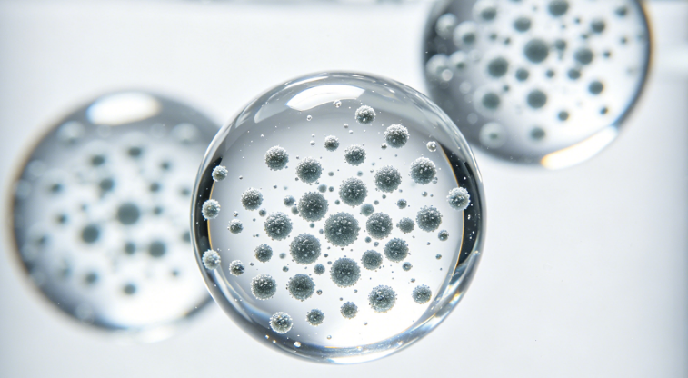
Antibody‑Conjugated Magnetic Bead Testing – Conjugation Efficiency, Activity, Stability, and Quality Control
If you are searching for antibody‑conjugated magnetic bead testing, you likely need to verify the performance of your immunomagnetic beads used in cell isolation, immunoprecipitation, exosome capture, lateral flow assays, or magnetic particle‑based diagnostics. Critical parameters include antibody loading density, binding activity, non‑specific binding (NSB), magnetic responsiveness, colloidal stability, and batch‑to‑batch reproducibility. Our laboratory provides comprehensive characterization of antibody‑conjugated magnetic beads – from routine protein quantification to flow cytometry‑based activity assays, dynamic light scattering (DLS) for aggregation, and functional capture efficiency testing in complex matrices – following ISO 13099 and ASTM standards for magnetic particles.
What We Analyze – Full Testing Scope for Antibody‑Conjugated Magnetic Beads
We do not simply report “antibody present”. Our platform includes total and surface antibody quantification by bicinchoninic acid (BCA) assay after bead digestion, orthogonal to fluorescent labeling (e.g., FITC‑anti‑IgG) and flow cytometry. For orientation and active site accessibility, we use Fab‑specific ELISA or a target antigen binding assay that measures only functional antibody (not denatured or buried). We perform binding capacity assays using purified antigen or target cells: we incubate beads with increasing amounts of antigen, wash, elute, and quantify captured antigen by LC‑MS/MS, ELISA, or fluorescence, determining maximum binding capacity (µg antigen/mg beads). Non‑specific binding (NSB) is assessed using irrelevant proteins (e.g., BSA, casein, or scrambled peptide) or negative cell lines, reported as % NSB relative to specific binding. For colloidal stability and aggregation, we use dynamic light scattering (DLS) and zeta potential measurement under storage and assay buffer conditions (pH, ionic strength, serum). Magnetic responsiveness is measured by sedimentation rate in a magnetic field (using a magnet and spectrophotometer), reporting capture half‑time (t50). Leaching of antibody is quantified by incubating beads in relevant buffer at 37°C for 24‑72h, then measuring released antibody by ELISA or BCA with detection limit 0.1 µg/mL. We also perform sterility and endotoxin testing (LAL assay) for IVD or therapeutic‑grade beads, and accelerated stability (40°C/75% RH for 1‑3 months) to predict shelf life.
Key parameters we routinely measure:
- Antibody loading (µg antibody/mg beads, or number of antibodies per bead) – CV <5% between replicates.
- Active antibody fraction (functional binding capacity / total loaded) – typically 50‑95% depending on conjugation chemistry.
- Binding capacity for target antigen (ng or µg per mg beads) – from Langmuir isotherm fitting.
- Non‑specific binding (NSB) to irrelevant proteins or cells – LOQ 0.1% of specific binding.
- Particle size distribution (mean diameter, polydispersity index PDI) – DLS, range 0.05‑10 µm.
- Zeta potential (surface charge, mV) – predictive of colloidal stability.
- Magnetic capture kinetics (t50 and % recovery) – using standard magnetic separator.
- Antibody leaching (released antibody under storage/assay conditions) – reported as ng/mg beads or % of total.
- Endotoxin (EU/mg beads) and bioburden – for in vivo or diagnostic applications.
- Residual blocking agent (BSA, casein, etc.) – by SDS‑PAGE or BCA after elution.
- Batch‑to‑batch reproducibility of all parameters – statistical analysis (mean, SD, %CV).
How Deep We Go – Single‑Bead Activity Analysis, In‑Process Capture Efficiency, and Long‑Term Stability Modeling
Most routine bead testing labs only measure total protein loading by BCA, which cannot distinguish inactive aggregates or buried antibodies. We provide single‑bead flow cytometry using fluorescent antigen to measure binding activity distribution across thousands of individual beads, identifying subpopulations with poor activity. For conjugation chemistry evaluation, we use peptide mapping (LC‑MS/MS) of trypsin‑digested beads to identify the site of conjugation (lysine, cysteine, carbohydrate) and quantify % of antibody molecules modified at each site. We also perform surface plasmon resonance (SPR) on beads captured on a sensor chip to directly measure real‑time binding kinetics (KD, kon, koff) of bead‑immobilized antibodies – an essential quality attribute for affinity applications. For complex matrix testing (serum, whole blood, cell lysates), we quantify capture efficiency and specificity by spiking target antigen into the matrix and measuring recovery after magnetic separation, plus evaluating co‑capture of abundant matrix proteins by LC‑MS/MS. Our stability studies go beyond simple leaching: we use DLS and activity assays weekly for up to 6 months at 4°C, 25°C, and 37°C, applying Arrhenius modeling to predict shelf life at recommended storage conditions. For sterile or clinical‑grade beads, we perform mammalian cell viability assays (MTT) on bead eluates to detect cytotoxic leachables.
Advanced capabilities include:
- Atomic force microscopy (AFM) imaging of individual beads – measure coating morphology and antibody layer thickness.
- Mass photometry for antibody density distribution – single‑molecule counting on bead surfaces.
- Competitive binding assays for epitope integrity – using labeled reference antibody and antigen.
- Magnetic particle spectrometer (MPS) for dynamic magnetic relaxation – relate to bead clustering in biological matrices.
- Simulated use‑cycle testing (capture‑wash‑elute, 10‑50 cycles) – measure degradation in activity and antibody loss.
- Protease resistance (incubation with trypsin or serum proteases) – remaining activity by ELISA.
- Matrix lot‑to‑lot variability assessment (e.g., different serum batches) – for diagnostic kit validation.
We routinely achieve measurement uncertainties: antibody loading ±5% (BCA), binding capacity ±8% (ELISA), NSB ±0.5% absolute, DLS PDI ±0.02, endotoxin <0.1 EU/mg. All methods follow ISO 13099 (Colloidal systems – zeta potential), ASTM E3264 (Magnetic particle characterization), and FDA guidance for immunomagnetic separation devices.
Why Choose Our Antibody‑Conjugated Magnetic Bead Testing – Key Advantages
1. ISO/IEC 17025:2017 accredited and GLP‑compliant workflows – suitable for R&D, QC, and regulatory submissions for diagnostic or therapeutic magnetic particles.
2. Orthogonal activity assessment (flow cytometry, SPR, cell binding, antigen ELISA) – we cross‑validate using multiple independent methods to ensure true functional activity, not just presence.
3. Single‑bead activity distribution (flow cytometry) – reveals batch heterogeneity that bulk assays miss, critical for consistent assay performance.
4. Ultra‑low detection of antibody leaching (down to 0.1 ng/µL by ELISA or MS) – essential for applications where leached antibodies cause background or toxicity.
5. Real‑world matrix testing (serum, plasma, whole blood, cell lysates) – we simulate your actual use conditions to report true capture efficiency and NSB.
6. Shelf‑life prediction via accelerated stability modeling (Arrhenius) – we save you months of real‑time waiting by predicting long‑term stability from high‑temperature data.
7. Fast turnaround with full data transparency – routine panel (loading, activity, NSB, size, zeta, leaching) in 5‑7 business days; full stability study in 4‑6 weeks (real‑time), 3 weeks (accelerated). You receive raw binding curves, DLS histograms, flow cytometry files, and detailed reports.
8. Custom method development for novel conjugation chemistries or bead types – click chemistry, streptavidin‑biotin, or custom coupling – we develop validated assays within 3‑4 weeks.
9. Competitive pricing for complete bead characterization packages – bundling loading, activity, NSB, leaching, size, zeta, and endotoxin costs 35% less than separate tests.
We have successfully completed over 300 antibody‑conjugated magnetic bead projects for diagnostic manufacturers, biotech companies, and academic research labs. Our team includes PhD biochemists and materials scientists specialized in magnetic particle functionalization and quality control.
Ready to Test Your Antibody‑Conjugated Magnetic Beads?
Provide your bead type (magnetic core, size, coating chemistry), antibody target, intended application (cell isolation, immunoassay, exosome capture), and critical quality attributes. We will provide a free technical consultation, a recommended testing strategy, and a fixed‑price quote. Whether you need QC release testing, stability validation, or troubleshooting of a failed batch, we deliver deep, accurate, and application‑relevant antibody‑conjugated magnetic bead testing tailored to your needs.