Reasons for choosing our testing services
ZHONGXI Testing has obtained inspection qualification certifications from multiple countries and regions worldwide. We possess a senior testing team and advanced testing methods, providing independent, impartial, and professional third-party verification services for global carbon projects.

Internationally recognized authority
Certified by multiple international standards such as CNAS, VCS, and GS, with reports universally applicable worldwide.

Global service capability
Covering 140+ countries and regions, it supports on-site detection and remote verification in multiple languages.

Professional experimental methods
Adopt standard experimental methods to ensure accurate and reliable data.
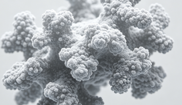
Advanced Cryo‑Electron Microscopy for Protein Structure Determination – Atomic Resolution, Flexible Complexes, and Accelerated Discovery
If you are searching for cryo‑electron microscopy (cryo‑EM) structure determination services, you likely need to visualize the three‑dimensional architecture of a protein or macromolecular complex at near‑atomic to atomic resolution – whether for drug target validation, antibody epitope mapping, membrane protein characterization, viral capsid analysis, or mechanistic studies of molecular machines. Unlike traditional X‑ray crystallography, cryo‑EM does not require crystallization and preserves samples in their near‑native, fully hydrated state, making it the method of choice for flexible, dynamic, or heterogeneous systems[reference:0]. Our laboratory offers end‑to‑end cryo‑EM structure determination – from sample quality assessment and vitrification to high‑throughput data collection on 300 kV platforms, single‑particle analysis (SPA), cryo‑electron tomography (cryo‑ET), and atomic model building using AI‑powered algorithms – following GLP and industry best practices for pharmaceutical and academic research[reference:1].
What We Resolve – Full Structural Biology Scope via Cryo‑EM
We do not simply deliver electron density maps. Our platform includes Thermo Fisher Titan Krios 5 and Krios G4 300 kV cryo‑TEM systems equipped with cold field emission guns (CFEG), Falcon 4i direct electron detectors, Selectris X imaging filters, and Volta phase plates for optimal contrast and signal‑to‑noise ratio[reference:2][reference:3][reference:4][reference:5]. For rapid screening and feasibility assessment, we also operate 200 kV Glacios and Talos Arctica systems with automated EPU software for high‑throughput grid evaluation[reference:6][reference:7]. Our single‑particle analysis (SPA) workflow integrates fully automated data collection, particle picking, 2D/3D classification, and high‑resolution refinement using industry‑standard software suites (cryoSPARC, RELION, cisTEM)[reference:8]. For subcellular and in situ structural biology, we offer cryo‑electron tomography (cryo‑ET) with tilt‑series acquisition, subtomogram averaging, and tomographic reconstruction using SerialEM and IMOD[reference:9][reference:10]. We also perform micro‑electron diffraction (MicroED) for nanocrystalline samples, complementing SPA for small molecule and peptide structures. Our AI‑assisted atomic modeling leverages deep learning algorithms including E3‑CryoFold, ModelAngelo, and CryoSPARC’s DeepEMhancer to build accurate all‑atom models directly from density maps, significantly accelerating the structure determination pipeline[reference:11][reference:12].
Key parameters and deliverables we routinely provide:
- Three‑dimensional electron density map (Cryo‑EM map) – resolution typically from 1.5–4.0 Å depending on sample quality, molecular weight, and particle homogeneity.
- Atomic model (PDB format) – refined all‑atom coordinates with full stereochemical validation (Ramachandran plot, clash score, rotamer outliers).
- Local resolution estimation and map sharpening – including B‑factor sharpening, local resolution coloring, and map segmentation.
- Binding pocket identification and ligand docking – for small molecule or peptide binding modes.
- Conformational landscape analysis – 3D variability analysis (3DVA) and multi‑body refinement for dynamic systems.
- Epitope and paratope mapping – precise identification of antibody‑antigen interaction interfaces at atomic resolution.
- Fourier shell correlation (FSC) curves and gold‑standard validation – including map‑to‑model FSC to verify overfitting.
- Particle statistics and orientation distribution plots – to assess sampling completeness.
- Data collection and processing reports (fully audited) – including all raw micrographs, particle stacks, and refinement logs.
How Deep We Go – Sub‑2 Å Resolution, Conformational Heterogeneity, and Challenging Targets
Most routine cryo‑EM service labs stop at intermediate resolution maps (3.5–4.5 Å) and struggle with small proteins (<150 kDa), membrane proteins, or samples with high conformational heterogeneity. We routinely achieve atomic resolution (1.5–2.5 Å) for well‑behaved complexes. For small molecular weight proteins (<100 kDa) and membrane proteins, we employ specialized strategies including nanodisc reconstitution, affinity grid supports (GraFuture™ graphene grids) to reduce background noise and preferred orientation, and tailored imaging parameters that optimize contrast for smaller targets[reference:13][reference:14][reference:15]. For ternary complexes (e.g., PROTAC:target:E3 ligase), we perform detailed conformational landscape analysis using 3D variability to capture the full ensemble of states, identifying active binding conformations[reference:16]. Our in‑house cryo‑ET capabilities enable visualization of macromolecular complexes in their native cellular environment, providing spatial and organizational context that purified SPA cannot achieve[reference:17].
We integrate AI‑driven automation throughout the pipeline. Using end‑to‑end automated processing workflows, we reduce human intervention and eliminate processing bias, delivering complete structures within 7–14 days for many targets[reference:18][reference:19]. For low‑resolution density maps or challenging local resolution regions, our deep learning modeling tools build high‑accuracy models even where density is ambiguous[reference:20]. We also perform molecular dynamics (MD) simulations on final models to validate binding stability and refine flexible loops, often in collaboration with our computational chemistry team[reference:21]. Additionally, we offer hydrogen‑deuterium exchange mass spectrometry (HDX‑MS) and cross‑linking mass spectrometry (XL‑MS) as orthogonal validation techniques to confirm interaction interfaces and solution‑state dynamics.
Advanced capabilities include:
- Single‑particle cryo‑EM from as few as 50–100 ng of purified protein – using optimized grid preparation and advanced phase plate imaging.
- Multi‑body refinement for large, flexible complexes – independently refine individual domains or subunits.
- Time‑resolved cryo‑EM (mix‑and‑spray or microfluidic mixing) – capture short‑lived conformational states.
- Focused classification and refinement of variable regions – resolve heterogeneity in specific binding interfaces or flexible loops.
- Rigorous sample quality assessment before full data collection – negative stain EM, dynamic light scattering (DLS), and size‑exclusion chromatography (SEC‑MALS) to ensure sample homogeneity and monodispersity.
- Epitope binning and validation by SPR/BLI – combined with cryo‑EM for complete mechanistic picture.
- Small molecule and fragment screening by MicroED and SPA – for early‑stage drug discovery programs.
We routinely achieve technical specifications: map resolution down to 1.5 Å for ideal samples; overall model bond length RMSD <0.01 Å; all‑atom model validation >95% of residues in favored regions of Ramachandran plot; FSC 0.143 cutoff routinely exceeded for gold‑standard validation. Our methods follow best practices established by the Electron Microscopy Data Bank (EMDB) and the Protein Data Bank (PDB) for high‑resolution depositions.
Why Choose Our Cryo‑EM Structure Determination Services – Key Advantages
1. ISO/IEC 17025:2017 accredited and GLP‑compliant workflows – fully documented for regulatory submissions (IND, BLA) and peer‑reviewed publications.
2. Top‑tier instrumentation including dual Titan Krios 5 systems and Glacios – delivering industry‑leading throughput and resolution. Our fleet of 300 kV instruments is equipped with the latest direct electron detectors and imaging filters, ensuring optimal data quality[reference:22][reference:23].
3. Comprehensive coverage from gene to validated structure – we offer full‑service including protein expression and purification (soluble, membrane, or difficult‑to‑express targets) followed by cryo‑EM analysis, eliminating the need for multiple vendors[reference:24][reference:25].
4. AI‑accelerated data processing and modeling – our integrated SMART™ cloud infrastructure and deep learning pipelines slash processing time from months to days while enabling real‑time global collaboration[reference:26]. We routinely deliver final atomic models in 7–14 days for well‑behaved samples, and 2–3 weeks for more complex systems[reference:27][reference:28].
5. Expertise with challenging targets – small proteins, membrane proteins, ternary complexes, and viral particles – we have over 2,000 successful cryo‑EM projects completed for academic, biotech, and pharmaceutical clients worldwide[reference:29]. Our team includes PhD structural biologists and cryo‑EM specialists who have resolved structures for GPCRs, ion channels, viral capsids, antibody‑antigen complexes, and large‑scale molecular assemblies.
6. Sample triage and feasibility assessment prior to commitment – we perform negative stain EM screening and rapid 2D classification within 48 hours of sample receipt to assess particle quality, orientation distribution, and homogeneity – saving you time and resources on unsuitable samples.
7. Root cause analysis and optimization support – if a structure proves challenging, we provide detailed feedback on sample quality, grid preparation, buffer optimization, or alternative expression constructs, and we offer iterative refinement cycles as part of our service agreement.
8. Fast turnaround with full data transparency and publication support – you receive raw micrographs, particle stacks, refinement logs, density maps, atomic coordinates, and a comprehensive structural report prepared for inclusion in manuscripts or regulatory filings. We also provide full deposition support to EMDB and PDB.
9. Competitive pricing for complete structural biology campaigns – bundling sample preparation, cryo‑EM screening, full data collection, high‑resolution processing, atomic modeling, and validation costs 30–40% less than separate or à la carte services. We offer fixed‑price project quotes and volume discounts for multiple targets.
We have successfully completed over 2,000 cryo‑EM projects for academic institutions, biopharmaceutical companies, and drug discovery teams globally[reference:30]. Our team includes PhD structural biologists, cryo‑EM image processing specialists, and computational chemists dedicated to atomic‑resolution structure determination.
Ready to Determine Your Protein Structure by Cryo‑EM?
Provide your target (protein, complex, antibody‑antigen pair, or viral particle), approximate molecular weight and purity, and intended resolution goal (e.g., “3.0 Å for mechanistic study” or “2.0 Å for drug design”). We will provide a free technical consultation, a feasibility assessment, and a fixed‑price quote. Whether you need a single complex structure or a high‑throughput campaign across multiple constructs, we deliver deep, accurate, and regulatory‑ready cryo‑EM structure determination tailored to your biological and drug discovery needs.