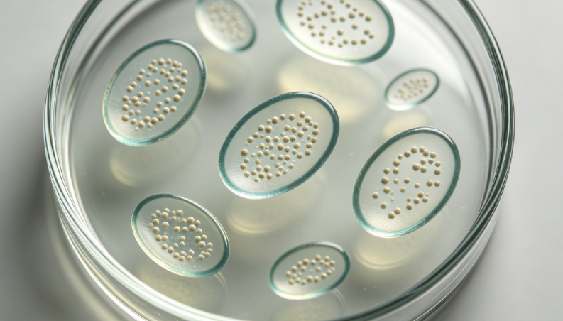
Reasons for choosing our testing services
ZHONGXI Testing has obtained inspection qualification certifications from multiple countries and regions worldwide. We possess a senior testing team and advanced testing methods, providing independent, impartial, and professional third-party verification services for global carbon projects.

Internationally recognized authority
Certified by multiple international standards such as CNAS, VCS, and GS, with reports universally applicable worldwide.

Global service capability
Covering 140+ countries and regions, it supports on-site detection and remote verification in multiple languages.

Professional experimental methods
Adopt standard experimental methods to ensure accurate and reliable data.
Comprehensive Lysosome Detection Service – From Acidic Compartment Phenotyping to Full Functional Profiling
You are searching for lysosome detection because you need to perform this assay—whether to monitor lysosomal health in drug discovery, quantify lysosomal enzyme activity, measure lysosomal membrane permeabilisation (LMP), or validate lysosomal biogenesis in autophagy research. We provide a full‑spectrum detection service that delivers quantitative, multiparametric, and high‑content readouts of lysosomal status across cell lines, primary cells, and tissues.
What We Detect – Beyond Simple Acidic Compartment Staining
Our lysosome detection goes far from basic LysoTracker® or acridine orange staining. We simultaneously assess lysosomal pH (by ratiometric probes such as LysoSensor™ or pH‑dependent GFP fusion proteins), lysosomal enzyme activity (cathepsins B, D, L, and acid phosphatase) via fluorogenic or FRET substrates, lysosomal membrane integrity (by Galectin‑3 puncta or LAMP‑1/2 exposure assay), and lysosomal abundance (by LAMP‑1/LAMP‑2 immunofluorescence or Western blot). For advanced studies, we quantify lysosomal Ca²⁺ release (using GCaMP variants) and TFEB nuclear translocation as a readout of lysosomal stress and biogenesis.
How Deep We Go – High‑Content Imaging, Flow Cytometry & Functional Multi‑Omics
We don't just report “lysosomes present”. Using automated high‑content confocal imaging (Opera Phenix®), we quantify per‑cell lysosome number, volume, circularity, spatial distribution, and co‑localisation with autophagosomes (LC3) or endosomes (Rab5/7) – processing up to 100,000 cells per sample with machine‑learning segmentation (CellProfiler/StarDist). For live‑cell dynamics, we perform time‑lapse lysosome pH tracking and lysosomal motility analysis (tracking speed, net displacement). Our spectral flow cytometry (Cytek® Aurora) resolves 11 lysosomal parameters simultaneously in heterogeneous populations, detecting even small subpopulations (as low as 0.5%) with distinct lysosomal phenotypes. For biochemical depth, we offer lysosomal immunoprecipitation (Lyso‑IP) followed by LC‑MS/MS proteomics to identify >1,500 lysosomal membrane and lumenal proteins from as few as 2×10⁶ cells.
Why Our Lysosome Detection Stands Out – Multiplexity, Sensitivity & Disease Relevance
1. Orthogonal validation: We never rely on a single dye. Each sample is cross‑checked with LAMP‑1/LAMP‑2 immunofluorescence, LysoTracker® Deep Red, and magic red cathepsin activity assay – ensuring true lysosomal signal versus autofluorescent or non‑specific compartments.
2. High‑throughput ready: Our 384‑well format supports siRNA or compound library screening (up to 50,000 wells/week). We provide Z'‑factor >0.6 for lysosomal pH assays, enabling robust hit identification.
3. Live‑cell & fixative compatibility: We optimise protocols for live‑cell imaging (37°C, 5% CO₂) as well as PFA‑fixed, FFPE tissue sections – including lysosomal antigen retrieval for archival samples.
4. Pathological reference database: Our internal atlas includes lysosomal fingerprints from 22 lysosomal storage disorders (LSDs) – Gaucher, Niemann‑Pick, Tay‑Sachs, etc. – allowing rapid classification of unknown phenotypes.
5. End‑to‑end service: We take cells, frozen tissues, or biopsy punches, perform optimised staining, imaging/flow cytometry, data analysis, and an interpretative report with statistical comparisons against controls.
Who Relies on Our Lysosome Detection – Real‑World Applications
Pharmaceutical companies use our service to screen lysosomal stabilisers for neurodegenerative disease (Parkinson’s, Alzheimer’s). One client identified a lead compound that restored lysosomal pH from 4.6 to 5.8 (pathological acidification) and rescued cathepsin D activity by 80% – data that supported their IND filing. Academic autophagy labs send us ATG knockout cell lines; we revealed a compensatory lysosomal biogenesis via TFEB even when canonical autophagy was blocked. A biotechnology company validated their lysosomal enzyme replacement therapy (ERT) by demonstrating precise LAMP‑1 co‑localisation and enzyme activity in patient fibroblasts – previously unseen by conventional plate readers.
Ready to Run Your Lysosome Detection?
Send us live or fixed cells (≥5×10⁵), cryosections (5‑10 sections), or tissue lysates (≥200 µg). We will perform multiplex lysosomal phenotyping (abundance, pH, enzyme activity, membrane integrity) – or a fully custom panel – and deliver quantitative data + high‑resolution images within 5‑7 business days. Request a free consultation; we will design the optimal lysosomal assay for your model (cell line, primary neuron, liver tissue, or tumour biopsy).