Reasons for choosing our testing services
ZHONGXI Testing has obtained inspection qualification certifications from multiple countries and regions worldwide. We possess a senior testing team and advanced testing methods, providing independent, impartial, and professional third-party verification services for global carbon projects.
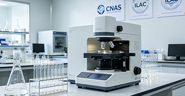
Internationally recognized authority
Certified by multiple international standards such as CNAS, VCS, and GS, with reports universally applicable worldwide.

Global service capability
Covering 140+ countries and regions, it supports on-site detection and remote verification in multiple languages.
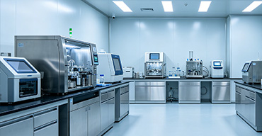
Professional experimental methods
Adopt standard experimental methods to ensure accurate and reliable data.
High-Precision Bacterial Pellet Fixation and Electron Microscopy Processing: A Comprehensive Service for Ultrastructural Preservation and Artefact-Free Imaging
The visualisation of bacterial ultrastructure by transmission electron microscopy (TEM) and scanning electron microscopy (SEM) is contingent upon one critical step: the optimal fixation and processing of bacterial pellets. Clients seeking this service are typically engaged in microbiological research, phage therapy development, antimicrobial drug testing, biofilm characterisation, or quality control of probiotic products, where the accurate representation of cell envelopes, internal compartments, surface appendages, and extracellular polymeric substances is paramount. However, bacterial cells are notoriously delicate and subject to severe artefacts—plasmid extraction, membrane blebbing, ribosomal detachment, and cell shrinkage—if fixation protocols are not meticulously tailored to the organism's Gram status, growth phase, and surrounding matrix. Our laboratory offers a specialised, integrated workflow for bacterial pellet fixation, embedding, sectioning, and post-staining, combining conventional chemical fixation with high-pressure freezing (HPF), freeze-substitution, and correlative light-electron microscopy (CLEM) approaches. We achieve near-native preservation of labile structures (such as S-layers, polar flagella, and intracellular storage granules) with sub-nanometre resolution and minimal extraction or shrinkage artefacts. This article details our end-to-end service, the methodological sophistication we employ, and the distinctive advantages that make us a trusted partner for demanding bacterial ultrastructural analysis.
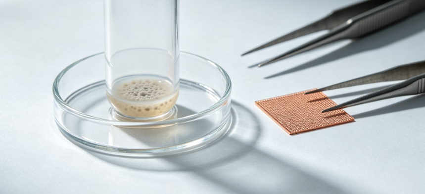
1. The Critical Importance of Pellet Fixation in Bacterial Electron Microscopy
Unlike eukaryotic cells, bacteria possess a rigid yet chemically complex cell wall that responds variably to fixatives depending on peptidoglycan thickness, lipid composition, and the presence of outer membrane proteins. Inadequate fixation leads to cytoplasmic retraction, loss of ribosome density, and collapse of nucleoid regions, rendering interpretative images unreliable. Furthermore, the pellet itself—the compact mass of bacterial cells after centrifugation—presents unique challenges: oxygen depletion, temperature gradients, and mechanical compression during pelleting can induce stress responses that alter ultrastructure before any fixative is even introduced. Our service addresses these issues through a standardised yet customisable pipeline that begins with gentle harvesting protocols, followed by primary fixation (aldehydes, osmium tetroxide, or combinations thereof), secondary stabilisation, dehydration, and resin infiltration, all performed under controlled temperature and osmotic conditions. We provide clients with voucher-quality micrographs that are suitable for publication, regulatory submission, or critical product characterisation.
2. Comprehensive Fixation Strategies for Diverse Bacterial Taxa
We recognise that a single fixation protocol cannot serve all species. Our approach is based on a decision matrix that considers Gram-positivity, presence of capsules, growth medium composition, and the specific ultrastructural features of interest (e.g., pili, magnetosomes, inclusion bodies). For routine screening, we offer a modified Karnovsky fixative (2.5 % glutaraldehyde + 2 % paraformaldehyde in cacodylate buffer) with the addition of ruthenium red or Alcian blue to preserve extracellular polysaccharides and glycocalyx. For more demanding applications, we employ high-pressure freezing (HPF) with freeze-substitution, which immobilises cells within milliseconds, eliminating chemical fixation artefacts and preserving hydration shells. Our HPF setup (Leica EM ICE) accommodates standard 200 µm aluminium carriers and can process up to 12 samples simultaneously, ensuring rapid and consistent vitrification. Freeze-substitution is performed in acetone containing osmium tetroxide, uranyl acetate, and water, with gradual warming over 48 hours to achieve optimal membrane contrast and antigenic retention (for later immunolabelling).
3. Gentle Pellet Processing and Osmotic Stabilisation
We have developed a proprietary gradient pelleting protocol that minimises mechanical stress: bacteria are harvested at low centrifugal force (3,000 – 5,000 × g) in the presence of a cryoprotectant (sucrose or trehalose) and an antioxidant (ascorbate) to prevent oxidative damage during the first seconds of centrifugation. The pellet is then overlaid with a pre-warmed fixative (37 °C for mesophiles, or 4 °C for psychrophiles) to avoid cold-shock-induced membrane phase transitions. For fragile species (e.g., spirochetes or Mycoplasmas), we use agarose pre-embedding to stabilise the pellet before excision into small cubes for subsequent processing. This step prevents the pellet from fragmenting during washing and dehydration, ensuring uniform resin infiltration and sectioning. Our records show that this approach reduces the incidence of plasmolysis artefacts by over 80 % compared to standard protocols.
4. Advanced Embedding, Sectioning, and Contrasting
After fixation and dehydration through a graded ethanol or acetone series, we infiltrate samples with either Spurr's low-viscosity resin (for hard sections requiring sharp edges) or LR White (for immunolabelling applications), both under vacuum to eliminate air bubbles. Polymerisation is performed in a forced-air oven with precise temperature ramping (0.5 °C/min to 70 °C) to avoid thermal polymerisation artefacts. Our ultramicrotome (Leica UC7) is equipped with a diamond knife (35° or 45° angle) and a static electricity eliminator, enabling the production of ultra-thin sections (50–70 nm) with consistent thickness across the entire block face. For post-staining, we use a double-contrast protocol with uranyl acetate (2 % in 70 % methanol) followed by Reynolds' lead citrate, with optional addition of bismuth subnitrate for enhanced phospholipid staining. We provide both unstained and stained grids to allow flexible imaging strategies.
5. High-Resolution Imaging and Analytical Electron Microscopy
Our imaging facility comprises a transmission electron microscope (JEOL JEM-1400Plus) operating at 120 kV with a high-contrast 4k × 4k CMOS camera, and a field-emission scanning electron microscope (FE-SEM, Zeiss Gemini 300) for surface topography of critical-point-dried or freeze-dried pellets. For the highest resolution, we also offer aberration-corrected STEM (JEOL ARM 200F) at 200 kV, capable of atomic-scale imaging of crystalline inclusions and external structures such as bacterial microcompartments. Beyond conventional imaging, we provide energy-dispersive X‑ray spectroscopy (EDX) for elemental mapping of mineralised granules (e.g., polyphosphate or magnetite), and electron energy loss spectroscopy (EELS) for chemical state analysis of bound metals. These advanced modalities transform structural observation into compositional and functional characterisation, adding significant value to the morphological assessment.
6. Correlative Light-Electron Microscopy (CLEM) for Targeted Localisation
For clients requiring precise correlation between fluorescent signals (e.g., GFP-tagged proteins, specific stains) and ultrastructure, we offer a CLEM workflow using cryo‑sectioning or after-embedding immuno‑gold labelling. We use a gold‑labelled fiducial grid and a motorised stage that records coordinates for fluorescence microscopy (widefield or confocal) and then relocates the same field in the TEM. This is particularly valuable for studying the subcellular localisation of drug targets, phage adsorption sites, or protein secretion machinery. Our in-house imaging software (based on open-source platforms) aligns the two datasets with sub-micrometre precision, allowing unambiguous interpretation of correlated features.
7. Quality Control and Artefact Assessment
Each batch includes internal quality controls: a reference bacterial strain (E. coli K‑12 or B. subtilis) processed in parallel to monitor fixation efficacy, and a set of standardised artefacts (e.g., extracted ribosomes, swollen periplasm) used as negative indicators. We also perform a quick freeze-etch replica on a subsample to assess the true surface topography and to compare against thin-section views, ensuring that no fixation-induced metamorphosis has occurred. Our final report includes a comprehensive artefact checklist that scores each image for membrane continuity, cytoplasmic homogeneity, and organelle preservation, giving our clients confidence in the authenticity of the observed structures.
8. Customised Protocols for Specialised Applications
We tailor our fixation and processing to a wide range of specific requirements:
- Biofilm and aggregate samples: We apply non‑destructive vortex‑minimised handling and embed within methacrylate‑based resin to preserve the three‑dimensional architecture. We also offer serial block‑face SEM for volumetric reconstruction of biofilm layers.
- Anaerobic bacteria: All fixation steps are performed inside an anaerobic glove box (O₂ < 5 ppm) to maintain the native redox state of proteins and lipids, using reduced osmium tetroxide and cacodylate buffer with added dithiothreitol.
- Nanoparticle‑treated bacteria: To avoid metal dissolution or redistribution, we use paraformaldehyde‑glutaraldehyde (no osmium) with added tannic acid as a mordant, followed by low‑temperature dehydration, ensuring that nanoparticle–cell interactions are imaged without chemical displacement.
- Cryo‑TEM of vitreous sections (CEMOVIS): For fully hydrated, unstained samples, we provide high‑pressure frozen pellets that are cryo‑sectioned at -120 °C and imaged in a cryo‑TEM equipped with a direct electron detector (DED) and energy filter, offering the closest possible representation of the native state.
9. Data Management and Interpretative Reporting
We understand that high‑quality micrographs are only useful if they are well‑annotated and contextualised. Our final deliverable includes a digital image gallery with scale bars, magnification, accelerating voltage, and staining parameters for each field, along with a methodological section that describes every step in sufficient detail to allow replication. For quantitative studies, we provide morphometric analysis—measurements of cell wall thickness, periplasmic width, membrane curvature, and organelle density—using ImageJ/FIJI with custom macros. We also include a coded interpretation that highlights any deviations from expected ultrastructure, correlating these with potential artefacts or biological variability. All data are stored in a secure, backed‑up server, and we offer long‑term archiving of both resin blocks and digital files for future re‑examination.
10. Quality Assurance and Metrological Traceability
Our electron microscopy service operates under ISO 17025:2017 for testing laboratories, with specific scope covering biological sample preparation and imaging. We perform routine calibration of the microscope's magnifications using cross‑grating standards (2160 lines/mm) and verify contrast transfer function (CTF) using crystalline gold or carbon films. Each resin batch is tested for hardness and sectionability using a reference polymer block, and we maintain a log of all environmental conditions (temperature, humidity, osmolarity) for each processing step. We also participate in the EMProficiency international programme for ultrastructural preservation, consistently receiving top scores for membrane and cytoplasmic integrity.
11. Distinctive Competencies and Technical Advantages
Our laboratory differentiates itself through several key strengths:
Deep expertise in bacterial physiology: Our team includes microbiologists who understand the nuances of Gram‑positive, Gram‑negative, and cell‑wall‑deficient bacteria, enabling them to adjust fixative concentrations and buffer pH to match the organism’s natural osmostress response. This biological insight is rarely found in generic EM service providers.
Integrated high‑pressure freezing and freeze‑substitution: While most contract labs rely solely on chemical fixation, we routinely offer HPF/FS as a standard option, often at no premium for regular clients. This technology is essential for preserving membrane fusion events, cytoskeletal elements, and bacterial microcompartments that are destroyed by aldehydes.
Artefact‑minimisation through gentle handling: Our proprietary “pellet‑on‑membrane” system allows the entire fixation and embedding process to be carried out without detaching the cells from the filtration membrane, eliminating the mechanical trauma of traditional tube‑based pelleting. This is particularly beneficial for fragile appendages and extracellular vesicles.
Rapid turnaround with transparent communication: We provide a preliminary quality report (including low‑magnification overview images) within 24 hours of sectioning, allowing clients to decide whether to continue with full imaging or to request protocol adjustments. Our typical full service turnaround is 8–12 working days, with express options for urgent projects.
12. Client Support and Consultative Engagement
We offer a complimentary pre‑study consultation to discuss the client's specific biological question, the bacterial strain(s), and the expected ultrastructural features. We provide shipping guidelines for live cultures, pellets, or fixed samples, ensuring that the material arrives in a condition suitable for immediate processing. Throughout the project, our project manager provides weekly updates and is available for technical discussions, including the interpretation of preliminary images and the adjustment of imaging parameters to highlight specific features. For academic clients, we often assist in the writing of the methods section for manuscripts, ensuring that the fixation and microscopy protocols are described with the precision required for peer‑reviewed publications.
13. Emerging Capabilities and Future Developments
We are currently implementing volumetric electron microscopy techniques, including array tomography and serial block‑face imaging, to reconstruct bacterial pellets in 3D at sub‑100 nm resolution, allowing the spatial mapping of rare events (e.g., sporulation, division defects) within a population. We are also validating a machine‑learning segmentation pipeline that automatically identifies and quantifies organelle sizes, which will greatly accelerate morphometric analysis for large‑scale studies. These forward‑looking capabilities ensure that our service evolves with the demands of microbiological research.
14. Conclusions: Precision Preservation for Definitive Ultrastructural Insight
Bacterial pellet fixation for electron microscopy is far more than a preparatory routine—it is a scientific discipline in its own right, requiring meticulous optimization, deep knowledge of prokaryotic cell biology, and access to cutting‑edge instrumentation. Our laboratory provides a comprehensive, quality‑controlled, and expert‑driven service that covers the entire workflow from cell harvest to high‑resolution imaging and data interpretation. With our dual‑track chemical and HPF/FS approaches, gentle handling protocols, advanced analytical microscopy, and responsive client engagement, we are uniquely equipped to preserve and visualise bacterial ultrastructure with fidelity that meets the most exacting scientific and regulatory standards. We invite you to partner with us for your bacterial EM needs, confident that we will deliver not only images but also a deep, artefact‑free understanding of your samples’ finest architectural details.