Reasons for choosing our testing services
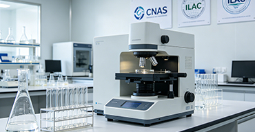
ZHONGXI Testing has obtained inspection qualification certifications from multiple countries and regions worldwide. We possess a senior testing team and advanced testing methods, providing independent, impartial, and professional third-party verification services for global carbon projects.
Internationally recognized authority
Certified by multiple international standards such as CNAS, VCS, and GS, with reports universally applicable worldwide.

Global service capability
Covering 140+ countries and regions, it supports on-site detection and remote verification in multiple languages.

Professional experimental methods
Adopt standard experimental methods to ensure accurate and reliable data.
Advanced Cytotoxicity Testing Services – From Regulatory‑Grade Screening to Mechanistic Profiling and High‑Content Analysis
The evaluation of Cytotoxicity is an essential component of the safety assessment of pharmaceuticals, medical devices, cosmetics, agrochemicals, and industrial chemicals. Determining the potential of a substance to cause cell death, inhibit proliferation, or induce sub‑lethal cellular damage is not only a regulatory requirement but also a critical step in the drug discovery process, biocompatibility testing, and product development. Regulatory agencies, including the FDA, EMA, and ISO (notably ISO 10993‑5), require that medical devices and their extracts be tested for Cytotoxicity using validated in vitro methods. Similarly, the ICH and USP guidelines mandate Cytotoxicity testing for pharmaceutical excipients, packaging materials, and biological products. However, a reliable Cytotoxicity assessment is not a simple “pass/fail” test; the outcome is highly dependent on the cell line selection, assay format, exposure conditions, detection method, and the interpretation of dose‑response data. A poorly designed assay may produce false‑negatives (due to compound interference, or insensitivity to specific modes of toxicity) or false‑positives (due to assay artefacts), leading to erroneous conclusions and potential regulatory setbacks. Our ISO/IEC 17025‑accredited and GMP‑compliant laboratory offers a fully integrated Cytotoxicity testing platform, encompassing a comprehensive suite of standardised and customised assays – from the basic metabolic activity and membrane integrity assays to advanced high‑content imaging, real‑time kinetic monitoring, and mechanistic pathway analysis. We deliver quantitative, traceable data, including IC50 values, full dose‑response curves, and in‑depth mechanistic insights, with expert interpretation that supports your product development and regulatory submissions.
1. The Scientific and Regulatory Rationale for Rigorous Cytotoxicity Testing
Cytotoxicity testing serves multiple critical purposes: (a) to establish the safety profile of a new chemical entity or material; (b) to rank the relative toxicity of candidate compounds or formulations; (c) to assess the biocompatibility of medical devices and their leachables (ISO 10993‑5); (d) to evaluate the potential for irritation or sensitisation; (e) to support the determination of safe exposure levels; and (f) to screen for mechanism‑specific toxicities (e.g., mitochondrial dysfunction, oxidative stress, or apoptosis). The choice of the assay system – whether metabolic (mitochondrial activity), membrane integrity (dye exclusion or LDH release), or morphological (microscopic evaluation) – must be aligned with the expected mechanism of toxicity, the nature of the test article (soluble, particulate, or extractable), and the regulatory requirements. A comprehensive Cytotoxicity study often involves multiple endpoints and time points to capture both immediate and delayed effects, and to distinguish between Cytotoxicity and cytostasis. Our service begins with a thorough consultation to select the most appropriate cell lines (e.g., L929, HeLa, HepG2, HaCaT, primary cells), exposure conditions, and endpoint assays, ensuring that the results are predictive of the in vivo response and compliant with the relevant guidelines.
2. Our Comprehensive Cytotoxicity Testing Platform – Multi‑Endpoint, Multi‑Technology
We maintain a complete suite of validated Cytotoxicity assays, all performed in a dedicated cell culture facility with stringent quality controls, and using well‑characterised cell lines (including ATCC and ECACC stocks) and certified reference materials (e.g., positive controls from the pharmacopoeias). Our portfolio includes:
2.1 Metabolic Activity Assays – MTT, XTT, WST‑1, and Resazurin (Alamar Blue)
We use tetrazolium reduction assays (MTT, XTT, WST‑1) and resazurin reduction to quantify the mitochondrial dehydrogenase activity of viable cells. We carefully optimise the incubation times and solubilisation steps to ensure linearity over a broad cell density range, and we include appropriate controls to correct for any direct interference by the test compound. The results are expressed as percentage viability relative to the untreated control, and we calculate the IC50 (the concentration causing 50% loss of viability) using four‑parameter logistic regression, providing a robust measure of potency.
2.2 Membrane Integrity Assays – Trypan Blue, Propidium Iodide, 7‑AAD, and LDH Release
We perform dye exclusion tests (trypan blue, PI, or 7‑AAD) to enumerate cells with compromised membranes, using either manual counting, automated image‑based cytometry, or flow cytometry. In parallel, we offer the lactate dehydrogenase (LDH) release assay, which measures the activity of this cytosolic enzyme in the culture medium following membrane damage. The LDH assay is particularly suitable for high‑throughput screening and for assays where test compounds interfere with metabolic dyes. We express the results as percentage Cytotoxicity relative to the maximum release control (using a lysis buffer).
2.3 Real‑Time Impedance‑Based Assays (xCELLigence, etc.)
We use label‑free, real‑time cell analysers to monitor cell viability and proliferation continuously by measuring the electrical impedance of the cell monolayer. This provides a kinetic profile of the cellular response, revealing the onset and duration of the cytotoxic effect, as well as any recovery phenomena. We derive multiple parameters: the cell index (CI), the time‑dependent IC50, and the slope of the response curve, which can indicate the rate of toxicity. This method is ideal for assessing compounds with rapid or delayed effects.
2.4 High‑Content Imaging (HCI) – Multiparametric, Mechanistic Cytotoxicity
We perform high‑content imaging using automated fluorescence microscopy (e.g., Operetta, ImageXpress) to acquire multi‑channel images and extract quantitative features at the single‑cell level. Our validated HCI panels include markers for: cell number and nuclear morphology (DAPI/Hoechst), mitochondrial membrane potential (JC‑1 or TMRM), reactive oxygen species (ROS) production (H2DCFDA), caspase‑3/7 activation (specific fluorescent substrates), and DNA damage (γH2AX). We quantify the percentage of cells exhibiting each feature and construct dose‑response curves for each parameter, providing a comprehensive mechanistic fingerprint of the toxicant. This approach is particularly powerful for distinguishing between apoptosis, necrosis, autophagy, and other modes of cell death.
2.5 Apoptosis and Necrosis Dual Staining – Annexin V / PI Assay
We use Annexin V‑FITC / PI staining to discriminate between viable, early apoptotic, late apoptotic, and necrotic cells. This assay is performed by flow cytometry or image‑based cytometry, and we report the percentage of cells in each quadrant. We can also assess the kinetics of apoptosis by performing this assay at multiple time points, providing insight into the mode of cell death and the involvement of specific caspases.
2.6 ATP Bioluminescence – CellTiter‑Glo® and Similar Assays
We use ATP bioluminescence to quantify the total ATP content, which is a sensitive indicator of metabolically active cells. The assay provides a wide linear range and is compatible with high‑throughput screening. We include an ATP standard curve to convert luminescence into absolute ATP concentration (pmol), enabling comparisons across experiments. This method is especially suitable for compounds that affect energy metabolism.
2.7 Clonogenic Survival Assay – Long‑Term Reproductive Integrity
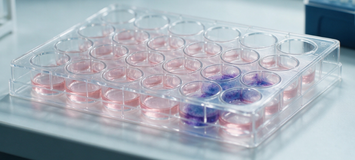
For evaluating the long‑term effects of test compounds, particularly those that cause DNA damage or chronic toxicity, we perform the colony formation assay. Cells are treated, then plated at low density and allowed to grow for 1‑3 weeks. The colonies (containing ≥ 50 cells) are fixed, stained, and counted. We express the results as surviving fraction relative to the control, which reflects the reproductive capacity of the cells and provides a measure of Cytotoxicity that is more relevant for certain endpoints (e.g., genotoxicity).
3. Advanced Capabilities – Mechanistic Deep‑Dive, Custom Models, and High‑Throughput Screening
For clients requiring a deeper understanding of the mode of action or a high‑level validation of their test system, we offer a suite of customised, research‑grade services that go beyond routine Cytotoxicity determination:
3.1 Comprehensive Mechanistic Panel – Apoptosis, Necroptosis, Autophagy, and Pyroptosis
We have developed a multi‑parameter panel that simultaneously probes multiple cell death pathways using a combination of fluorescent probes and immunofluorescence markers. This includes: (i) Caspase‑3/7 activation (apoptosis); (ii) RIPK1/RIPK3 phosphorylation (necroptosis); (iii) LC3‑II accumulation (autophagy); and (iv) IL‑1β and caspase‑1 activation (pyroptosis). The panel is applied on both adherent and suspension cell lines, and the data are analysed using automated image analysis to provide a quantitative profile of the dominant cell death pathway induced by your compound.
3.2 3D Culture and Organoid Cytotoxicity – Spheroid and Tumour Model Testing
We have adapted our Cytotoxicity assays to 3D spheroids and organoid cultures, which better mimic the in vivo microenvironment and provide more predictive toxicological data. We assess cell viability using ATP‑based assays, fluorescent live/dead staining, and confocal microscopy to evaluate the penetration of the compound into the spheroid and the distribution of cytotoxic effects. This service is particularly valuable for oncology drug screening and for assessing the toxicity of compounds with limited tissue penetration.
3.3 High‑Throughput Screening (HTS) – Compound Library Evaluation
We operate automated HTS platforms (384‑well and 1536‑well formats) with liquid‑handling robots, high‑speed plate readers, and automated image analysers. We can screen thousands of compounds per day, generating full dose‑response curves and IC50 values. We incorporate rigorous quality controls (Z′ factor > 0.6, signal‑to‑noise ratio > 10) and provide a comprehensive hit selection report, including a statistical analysis of the screen performance and a list of confirmed hits.
3.4 Co‑Culture and Immuno‑Cytotoxicity – CAR‑T, NK, and ADCC Assays
We perform immune cell‑mediated Cytotoxicity assays to evaluate the killing of target cells by effector cells (e.g., CAR‑T cells, NK cells). Using a combination of luciferase‑based target killing (with luciferase‑expressing target cells) and flow cytometry (with cell‑specific markers), we quantify specific lysis and calculate the EC50 of the effector cells or the potency of the therapeutic antibody in antibody‑dependent cell‑mediated Cytotoxicity (ADCC).
3.5 Method Validation and Regulatory Compliance (ISO 10993‑5, ICH Q2(R1), USP)
We provide full method validation for each Cytotoxicity test, including the demonstration of: (a) specificity (minimal interference from the test article); (b) linearity (over the relevant concentration range); (c) accuracy (by spike‑and‑recovery experiments using a reference cytotoxic agent); (d) precision (intra‑assay and inter‑assay reproducibility); (e) limit of detection (LOD) and limit of quantification (LOQ) for the viability endpoints; and (f) robustness to variations in cell passage, seeding density, and incubation conditions. The validation report is prepared in a format suitable for inclusion in regulatory submissions (e.g., 510(k), IDE, NDA, or BLA).
3.6 Extractable and Leachable Cytotoxicity Testing (ISO 10993‑12)
We provide specialised testing for medical device extracts and leachables, following ISO 10993‑12 for the preparation of extracts. We test the extracts using both the direct contact and agar overlay methods, with appropriate positive and negative controls, to evaluate the potential Cytotoxicity of device materials under simulated clinical use conditions. We also assess the effect of different extraction vehicles (e.g., DMEM, saline, and ethanol) on the Cytotoxicity profile.
4. Data Integrity, Standardisation, and Comprehensive Reporting
All assays are conducted under a strict quality management system that includes the use of certified reference materials, validated cell banks, routine positive and negative controls, and participation in proficiency testing schemes (e.g., UK NEQAS). Our reports provide:
- Full method description – detailing the cell line, culture conditions, treatment protocol, assay type, and detection parameters.
- Detailed results – including dose‑response curves, IC50 values (with 95% confidence intervals), and, for kinetic assays, time‑course profiles.
- Validation data – summarising the linearity, precision, accuracy, and robustness of the assay.
- Graphical outputs – including representative dose‑response plots, microscopy images (phase contrast and fluorescence), and flow cytometry histograms.
- Interpretative comments – on the potential mechanism of toxicity, the relevance of the results to the intended use, and any recommendations for further testing.
Our reports are authored by senior toxicologists and cell biologists who provide not just raw data, but contextual interpretation to help you make informed decisions and to support your regulatory submissions.
Turnaround times: 3‑5 working days for standard Cytotoxicity screening (e.g., MTT or LDH), 1‑2 weeks for comprehensive mechanistic panels, and 2‑3 weeks for full method validation. Express services are available for urgent projects.
5. Our Unmatched Expertise – Why Choose Our Laboratory for Cytotoxicity Testing
We are one of the few laboratories that hold ISO/IEC 17025 accreditation and GMP compliance for Cytotoxicity testing across a broad spectrum of test articles (chemicals, pharmaceuticals, medical devices, and cosmetics). Our integrated infrastructure combines a state‑of‑the‑art cell culture facility, advanced imaging platforms, flow cytometry, impedance‑based analysers, and high‑throughput screening systems – enabling multi‑endpoint, multi‑technology cross‑validation and the ability to handle complex or unusual sample types. Our team comprises PhD‑level toxicologists, cell biologists, and regulatory specialists with extensive experience in assay development, method validation, and regulatory submissions, and we have a proven track record of supporting successful product registrations with the FDA, EMA, and other agencies.
We offer unparalleled flexibility: we work with a wide range of cell lines (including primary and stem cells) and can develop custom Cytotoxicity assays for your specific cell type, compound, or application. We provide customised sampling kits and guidance on sample handling and extraction procedures. Our secure online portal enables real‑time tracking, access to raw data, and downloadable certificates of analysis.
Our modular pricing ensures cost‑effectiveness: you select the level of analysis – from a simple screening to a full mechanistic and validation package – with discounts for routine monitoring programmes and bulk submissions. We also provide training, method transfer, and technical consultancy to help you build or strengthen your internal Cytotoxicity testing capabilities.
With a proven history of serving over 100 clients in the pharmaceutical, biotechnology, medical device, and cosmetic industries, we have enabled our partners to ensure product safety, achieve regulatory compliance, and accelerate time‑to‑market. We invite you to leverage our expertise for your Cytotoxicity testing needs.
Contact our technical experts today to discuss your test article, cell system, and specific regulatory or research requirements. We will design a tailored testing plan and provide a detailed proposal within one business day.