Reasons for choosing our testing services
ZHONGXI Testing has obtained inspection qualification certifications from multiple countries and regions worldwide. We possess a senior testing team and advanced testing methods, providing independent, impartial, and professional third-party verification services for global carbon projects.
Internationally recognized authority
Certified by multiple international standards such as CNAS, VCS, and GS, with reports universally applicable worldwide.

Global service capability
Covering 140+ countries and regions, it supports on-site detection and remote verification in multiple languages.
Professional experimental methods
Adopt standard experimental methods to ensure accurate and reliable data.
Bismuth Oxychloride (BiOCl) Nanosheet Characterization: Advanced Analytical Services for 2D Photocatalytic Materials
Bismuth oxychloride (BiOCl) nanosheets represent a burgeoning class of two-dimensional (2D) semiconductor materials that have attracted substantial research interest due to their unique layered structure, anisotropic physicochemical properties, and superior photocatalytic performance. The distinctive {001} facet exposure in BiOCl nanosheets generates an internal electric field perpendicular to the basal plane, which drastically promotes charge carrier separation and migration, making these 2D structures far more efficient than their bulk counterparts for applications in environmental remediation, water splitting, and antibacterial coatings. Clients seeking analytical services for BiOCl nanosheets are typically engaged in the synthesis, optimization, and quality assurance of these advanced materials for both academic research and industrial scale-up. The primary objectives of such testing are to verify the nanosheet morphology, thickness uniformity, lateral dimensions, crystallographic orientation, surface defect chemistry, optical band structure, and, most critically, the photocatalytic activity under relevant irradiation conditions. These parameters are highly sensitive to synthesis protocols—including hydrothermal temperature, precursor ratio, surfactant selection, and exfoliation efficiency—and cannot be reliably evaluated using bulk material characterization methods alone. This article presents a rigorous technical overview of our comprehensive testing capabilities for BiOCl nanosheets, encompassing state-of-the-art analytical techniques, metrological traceability, and expert data interpretation that transforms complex raw data into actionable insights for material development and process control.
Structural and Morphological Peculiarities of BiOCl Nanosheets and Their Testing Implications
BiOCl crystallizes in a tetragonal matlockite structure (space group P4/nmm), with strong intralayer covalent bonding within the [Bi₂O₂]²⁺ slabs and weak interlayer van der Waals forces between the chloride ion layers. This anisotropic bonding imparts a pronounced tendency for preferential growth along the (001) direction, yielding ultrathin nanosheets with large exposed basal planes. The thickness of these nanosheets, typically ranging from 1 to 20 nm, directly influences the quantum confinement effect, bandgap widening, and the density of surface oxygen vacancies. The lateral size (from tens of nanometers to several micrometers) affects the specific surface area, light scattering, and the diffusion path of photogenerated carriers. The exposed facet ratio, particularly the dominance of (001) over (110) or (010) facets, governs the internal electric field strength and the adsorption affinity for pollutant molecules. Furthermore, BiOCl nanosheets often contain a high density of oxygen vacancies and bismuth sub-lattice defects that serve as active sites for molecular oxygen activation and enhance visible-light absorption. These structural and morphological attributes are interdependent and require a suite of complementary techniques for accurate evaluation. Our testing program is specifically designed to capture these 2D-specific characteristics, employing methods that are sensitive to the vertical dimension, crystallographic texture, and surface chemistry at the nanometer scale—capabilities that conventional powder diffraction or bulk optical spectroscopy cannot provide.
Regulatory and Reference Frameworks for Nanosheet Characterization
Although no single standard exists exclusively for BiOCl nanosheets, our testing protocols are established in accordance with internationally recognized guidelines for nanomaterials and photocatalytic materials. For dimensional characterization, we adhere to the principles of ISO 21363:2020 (Nanotechnologies — Measurements of particle size and shape distributions by transmission electron microscopy) and ISO 19749:2021 (Nanotechnologies — Measurement of particle size and shape distributions by scanning electron microscopy). For photocatalytic performance, we follow ISO 10678:2010 for aqueous methylene blue degradation and ISO 22197-1:2016 for gas-phase NO removal, with appropriate modifications to accommodate the thin-film or powder form of the nanosheets. Additionally, we reference the guidance provided by the OECD Working Party on Manufactured Nanomaterials for the physicochemical characterization of nanomaterials in regulatory dossiers. Our measurements are performed under strictly controlled environmental conditions (temperature 25.0 ± 0.5 °C, relative humidity 50 ± 2%) and with light sources calibrated against NIST-traceable radiometric standards. We include reference materials—such as size-certified polystyrene nanospheres for microscopy and commercial TiO₂ (P25) for photocatalytic benchmarking—in each analytical batch to ensure reproducibility and enable cross-laboratory comparison. All our reports explicitly state the test conditions, the statistical treatments applied, and the measurement uncertainties, providing full traceability and defensibility for publication or regulatory filing.
Advanced Characterization Methodologies: Morphology, Thickness, and Lateral Dimensions
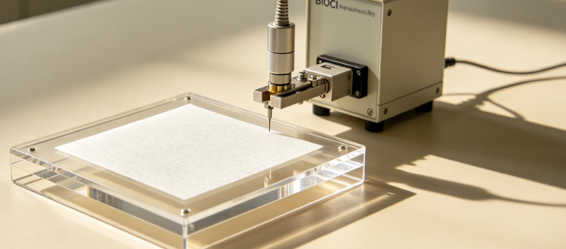
Accurate assessment of the 2D morphology of BiOCl nanosheets is the cornerstone of our testing service. We employ high-resolution transmission electron microscopy (HRTEM) operated at 200 kV with a field-emission gun, equipped with a high-angle annular dark-field (HAADF) detector for scanning TEM (STEM) mode. This allows for direct visualization of the lattice fringes corresponding to the (001) and (110) planes, from which we determine the crystal orientation and the presence of edge dislocations or stacking faults. For thickness measurement, we use atomic force microscopy (AFM) in tapping mode, scanning over an area of at least 5 µm × 5 µm with a vertical resolution of 0.03 nm, to obtain statistically meaningful thickness distributions (minimum 100 individual nanosheets). We also utilize electron energy loss spectroscopy (EELS) in the TEM to map the plasmon energy shift, which correlates with thickness at the sub-nanometer scale. Lateral dimensions (length and width) are determined from both SEM and TEM images using automated image analysis software, providing histograms of size distribution and aspect ratios. To complement these direct imaging techniques, we perform dynamic light scattering (DLS) and nanoparticle tracking analysis (NTA) for liquid-dispersed nanosheets to obtain the hydrodynamic diameter and the polydispersity index (PDI), which are essential for assessing colloidal stability and processability. For a more quantitative crystallographic texture, we conduct grazing-incidence X-ray diffraction (GIXRD) with a parallel-beam geometry and a 2D detector, allowing us to compute the Lotgering orientation factor for the (001) reflection and to assess the degree of preferred orientation in thin-film assemblies. This multi-technique morphological interrogation ensures that we capture both the intrinsic dimensions of individual nanosheets and the ensemble behavior of the powder or dispersion.
Surface Chemistry, Defect Engineering, and Electronic Property Assessment
The unique catalytic properties of BiOCl nanosheets are largely governed by their surface chemical state, particularly the concentration and distribution of oxygen vacancies (OVs) and bismuth-related defect states. We employ X-ray photoelectron spectroscopy (XPS) with monochromated Al Kα radiation and a hemispherical analyzer, performing high-resolution scans of Bi 4f, O 1s, and Cl 2p core levels. The O 1s spectrum is deconvoluted into lattice oxygen (typically at 529.8 eV), oxygen vacancies (≈531.2 eV), and chemisorbed hydroxyl or adsorbed water (≈532.5 eV), providing a quantitative OV density relative to the total oxygen signal. For a more sensitive detection of paramagnetic defect centers, we carry out electron paramagnetic resonance (EPR) spectroscopy at both room temperature and 77 K, using a microwave frequency of 9.5 GHz (X-band), to identify the signature of oxygen vacancies (g ≈ 2.001) and any possible bismuth sub-lattice defects. The optical properties are assessed by UV-Vis-NIR diffuse reflectance spectroscopy with an integrating sphere, and the bandgap is extracted via Tauc plots, with special attention to the indirect transition characteristic of BiOCl. To probe the electronic band structure in greater depth, we perform ultraviolet photoelectron spectroscopy (UPS) on spin-coated nanosheet films, using He I (21.22 eV) excitation, to determine the ionization potential (i.e., the valence band maximum relative to the vacuum level), and combined with the optical bandgap, we construct a complete energy band diagram that includes the conduction band position—a vital parameter for predicting the photocatalytic redox power. We also conduct Kelvin probe force microscopy (KPFM) on individual nanosheets to map the surface potential and work function variation, correlating these with local defect distributions and facet-dependent electronic properties. These comprehensive surface and electronic analyses provide a deep mechanistic understanding of the nanosheet's reactivity and offer clear guidance for defect engineering or surface passivation strategies.
Photocatalytic Performance Evaluation Tailored to Nanosheet Geometry
The photocatalytic activity of BiOCl nanosheets is evaluated using protocols specifically adapted to their 2D morphology, which affects light absorption, mass transport, and active site availability. Our standard assay employs methylene blue (MB) degradation in aqueous suspension under controlled stirring, with a fixed photocatalyst concentration (0.1 g/L) and a 300 W Xe lamp equipped with an AM 1.5G filter and a custom UV bandpass filter (300–400 nm) to match the absorption edge of pure BiOCl. For doped or sensitized nanosheets, we use a UV-cutoff filter (λ ≥ 420 nm) to isolate visible-light activity. The degradation kinetics are monitored by in-situ UV-Vis spectroscopy at 664 nm, and we compute the apparent first-order rate constant (k) via linear regression, with all measurements performed in triplicate. Beyond MB, we offer a panel of probe molecules, including rhodamine B (RhB), methyl orange (MO), and 4-chlorophenol (4-CP), to assess the versatility of the nanosheet catalyst. For applications in gas-phase purification, we conduct NO removal tests in a flow-through reactor, measuring NO and NO₂ concentrations with a chemiluminescence analyzer and calculating the NO conversion efficiency. To evaluate the photocatalytic stability, we perform cycling experiments (five to ten consecutive runs) and characterize the nanosheets by TEM and XPS post-test to detect any morphological or chemical changes. Additionally, we carry out scavenger experiments using tert-butanol (•OH quencher), benzoquinone (•O₂⁻ quencher), and EDTA (h⁺ quencher) to identify the dominant reactive oxygen species, providing mechanistic insights. Importantly, we normalize the photocatalytic activity by the effective surface area of the nanosheets (determined from BET analysis and geometric calculations) to obtain an intrinsic activity descriptor that allows fair comparison across samples with different sizes or thicknesses. This approach ensures that performance differences are attributed to true catalytic properties rather than to surface area variations.
Our Technical Capabilities: Precision, Sensitivity, and Diagnostic Resolution
Our laboratory is equipped with a state-of-the-art nanosheet characterization platform that integrates all required analytical modules in a controlled environment (class 1000 cleanroom for sample preparation). The HRTEM (FEI Titan Themis) operates at 80–300 kV and is equipped with a spherical aberration corrector for imaging and STEM, providing a point resolution of 0.08 nm and an energy resolution of 0.5 eV for EELS. The AFM (Bruker Dimension Icon) operates in PeakForce Tapping mode with ScanAsyst optimization, achieving a vertical resolution of 0.03 nm and a lateral resolution of 0.2 nm. The XPS/UPS system (PHI VersaProbe 4) uses a monochromatic Al Kα source (1486.6 eV) with a spot size down to 10 µm and a He I UV source for UPS, providing a binding energy resolution of 0.1 eV. The EPR spectrometer (Bruker EMXplus) operates at X-band with a sensitivity of 1 × 10⁹ spins/G and offers variable-temperature capability (4–300 K). For optical measurements, we use a UV-Vis-NIR spectrophotometer (PerkinElmer Lambda 1050+) equipped with a 150 mm integrating sphere and a PbS detector for the near-infrared range, with a photometric accuracy of ±0.003 absorbance units. The photocatalytic testing system includes a custom-designed photoreactor with temperature control, magnetic stirring, and a calibrated solar simulator (Oriel LCS-100) with an array of optical filters, and we monitor light intensity continuously with a NIST-traceable power meter. All instruments undergo annual calibration traceable to NIST and PTB, and we participate in interlaboratory comparisons for nanoparticle size and photocatalytic activity. Our quality management system follows ISO/IEC 17025 and ISO 9001, and we maintain a stringent data integrity protocol that records all environmental parameters and instrument settings for each measurement, enabling full reproducibility.
Our combined measurement precision allows us to report thickness with an uncertainty of ±0.2 nm (for AFM), lateral dimensions with ±3%, OV density via XPS with a relative standard deviation of ±2.5%, and photocatalytic rate constants with an expanded uncertainty (k=2) of ±4%. This level of precision is essential for detecting the subtle effects of synthesis variations—for example, a change in hydrothermal temperature of 5 °C often leads to a thickness change of 0.5 nm and a corresponding 15% change in activity, which our systems can reliably differentiate. We also provide correlative microscopy by identifying the same nanosheet region in AFM, SEM, and TEM, enabling cross-validated dimensional measurements.
Distinguishing Advantages of Our BiOCl Nanosheet Testing Service
Our service offers several unique advantages that directly address the specific challenges of 2D material characterization. First, we provide a fully integrated “nanosheet-to-performance” testing package that covers morphology, thickness, crystal structure, surface chemistry, electronic bands, and photocatalytic activity in a single workflow, eliminating the need for clients to coordinate multiple specialized facilities and reducing total turnaround time to 12–18 working days. Second, we employ a proprietary thickness-correlation algorithm that combines AFM, TEM, and optical reflectance data to produce a reliable thickness map for large-area nanosheet films, which is particularly valuable for clients developing coatings or membranes. Third, our team includes senior scientists with over 40 years of combined experience in layered materials and photocatalysis, providing exceptional interpretative expertise—we can distinguish between intrinsic activity, artifact effects (such as aggregation or light attenuation), and the influence of substrate interactions. Fourth, we offer customizable test matrices that can include additional techniques such as Raman spectroscopy for strain analysis, zeta potential for surface charge assessment, and time-resolved microwave conductivity for carrier mobility. We also provide accelerated aging tests under continuous UV or visible irradiation for up to 200 hours to evaluate photostability and phase stability, and we can perform in-situ experiments that monitor structural changes during photocatalytic reactions. Our clients also benefit from a secure web-based portal for real-time data access and collaborative interpretation sessions.
Typical Applications and Customization for Specific Use Cases
Our characterization services are adaptable to a broad spectrum of BiOCl nanosheet applications. In environmental remediation, we optimize nanosheets for the photodegradation of dyes, antibiotics, and persistent organic pollutants, with the option to include wastewater matrix spiking. For photocatalytic hydrogen production, we add headspace gas chromatography (GC) to quantify H₂ evolution rates and compute the apparent quantum yield (AQY). In self-cleaning coatings, we evaluate the nanosheet films for superhydrophilicity and oil-fouling removal under standard indoor lighting. For antibacterial surfaces, we perform bacterial viability assays (against E. coli and S. aureus) under relevant illumination conditions. We can also tailor the light source spectrum using a series of narrow-band LEDs (from 280 nm to 700 nm) to match specific application environments, such as UV-LED water purifiers or solar simulators for outdoor use. Furthermore, we offer variable-temperature photocatalytic testing (5–80 °C) to assess the thermal robustness of the nanosheets and to derive activation energies. Our team collaborates with clients to design a test plan that addresses their specific development questions—whether it is the effect of a new surfactant on nanosheet thickness, the impact of doping on defect chemistry, or the scalability of a synthesis route.
Case Example: Decoupling Thickness and Defect Contributions to Photocatalytic Activity
A recent project with an academic research group illustrates the diagnostic power of our integrated approach. The client had synthesized BiOCl nanosheets with three distinct thickness regimes (≈3 nm, ≈8 nm, and ≈15 nm) by varying the amount of a structure-directing agent. Preliminary optical and simple MB tests suggested that the thinnest nanosheets had the highest activity, but the client was uncertain whether this was due to confinement effects or the higher surface area. Our comprehensive analysis revealed that while the 3 nm nanosheets indeed had the widest bandgap (3.42 eV vs. 3.32 eV for the 15 nm ones), they also exhibited the highest oxygen vacancy density (OV/Bi ratio of 0.18 vs. 0.10). Using our correlation engine, we identified that the OV density was the dominant factor (R² = 0.92) in determining the rate constant, whereas thickness played a secondary role through the surface area contribution. We further performed KPFM mapping, which showed that the thinner nanosheets had a more negative surface potential, enhancing the electron transfer to adsorbed O₂. The client used these insights to optimize their synthesis to maximize OVs while preserving the thin morphology, achieving a 35% improvement in activity over their initial best sample. This case demonstrates that our testing not only quantifies properties but provides a mechanistic framework for rational design—a capability far beyond standard report generation.
Our Engagement Process and Client Support
To begin a project, clients submit a sample information form detailing the nanosheet synthesis method, intended application, and specific analytical priorities. We then provide a customized test plan and a fixed-price quotation, with a timeline agreed upon before any work commences. Upon sample receipt, we perform an initial quality check (visual and low-magnification imaging) to confirm the sample meets the minimum requirements for testing. We proceed with non-destructive characterization (AFM, SEM, DRS, XPS) before any destructive or consuming tests (e.g., photocatalytic cycling or TEM cross-sections). Throughout the project, we provide weekly progress updates and are always available for technical discussions. The final comprehensive report includes raw data in multiple formats (images, spectra, spreadsheets), processed results with detailed statistical analysis, a comparison to literature data for BiOCl nanosheets, and a summary of key findings and actionable recommendations. We also include a clear statement of measurement uncertainties and the corresponding standard references. We offer a confidentiality agreement to protect proprietary synthesis details, and we can arrange for priority processing (5–7 working days) at an additional fee. Volume discounts are available for multi-sample series, such as optimization arrays or batch-to-batch quality control.
Our commitment to scientific excellence, metrological traceability, and interpretive depth has established us as a trusted partner for researchers and developers working with BiOCl nanosheets. By choosing our service, you gain access to a team of dedicated experts who will treat your materials with the highest level of rigor and care. We invite you to contact our technical team for a complimentary consultation to discuss your specific requirements and to explore how our advanced characterization can accelerate your nanosheet development and ensure the reliability of your photocatalytic applications. We are confident that our comprehensive, insight-rich reporting will empower you to make informed decisions and achieve superior material performance.