Reasons for choosing our testing services
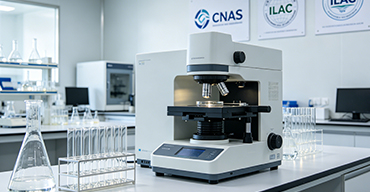
ZHONGXI Testing has obtained inspection qualification certifications from multiple countries and regions worldwide. We possess a senior testing team and advanced testing methods, providing independent, impartial, and professional third-party verification services for global carbon projects.
Internationally recognized authority
Certified by multiple international standards such as CNAS, VCS, and GS, with reports universally applicable worldwide.

Global service capability
Covering 140+ countries and regions, it supports on-site detection and remote verification in multiple languages.

Professional experimental methods
Adopt standard experimental methods to ensure accurate and reliable data.
Comprehensive Physicochemical and Functional Characterization of Magnetic Silica Nanospheres: Advanced Analytical Services for Biomedical, Environmental, and Catalytic Applications
Magnetic silica nanospheres (MSNS) – typically comprising a superparamagnetic iron oxide core (e.g., magnetite or maghemite) encapsulated within a mesoporous or dense silica shell – have become indispensable in diverse fields including targeted drug delivery, bioseparation, immunoassay, environmental remediation, and heterogeneous catalysis. Their performance is governed by a delicate interplay of nanoparticle size and size distribution, core/shell morphology, silica porosity and surface chemistry, magnetic saturation and coercivity, colloidal stability, and the density of surface functional groups (e.g., amino, carboxyl, thiol). Even minor batch‑to‑batch variations can drastically affect the binding capacity, separation efficiency, biocompatibility, and catalytic activity. Therefore, a rigorous, multi‑parametric analytical regime is essential for material development, quality assurance, process optimization, and regulatory compliance. Our ISO/IEC 17025‑accredited laboratory offers a dedicated, high‑depth testing service specifically tailored for magnetic silica nanospheres, integrating advanced microscopic, spectroscopic, magnetic, and thermal methods to deliver a complete physical, chemical, and functional fingerprint – far beyond routine SEM or dynamic light scattering (DLS) alone.
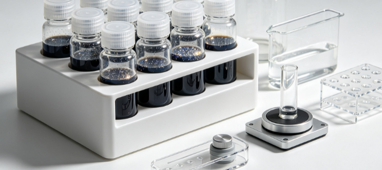
Why Comprehensive Characterization of Magnetic Silica Nanospheres Is Critical for Advanced Applications
Clients seeking our testing services typically aim to: (i) accurately determine the core diameter, shell thickness, and overall hydrodynamic size to ensure consistency with the intended application (e.g., cell separation requires narrow size distribution), (ii) quantify the magnetic properties (saturation magnetization, coercivity, remanence) to guarantee efficient separation under magnetic fields, (iii) measure the specific surface area, pore volume, and pore size distribution of the silica shell to predict drug loading or catalyst immobilization capacity, (iv) identify and quantify surface functional groups (e.g., –NH₂, –COOH, –SH) that govern the bioconjugation or adsorption selectivity, (v) evaluate the colloidal stability and zeta potential under various pH and ionic strength conditions to prevent aggregation in biological media, (vi) assess the iron leaching and shell integrity under acidic or enzymatic environments to ensure biocompatibility and safety, (vii) detect the presence of trace impurities or unreacted precursors that could cause toxicity or interference, and (viii) generate robust data for regulatory submissions, product certifications, and supplier qualification. Routine single‑parameter tests such as DLS or simple SQUID magnetometry are insufficient because the functional performance depends on the synergistic interplay of magnetic properties, pore architecture, surface chemistry, and colloidal behaviour, all of which require a suite of complementary techniques. Our integrated platform combines transmission electron microscopy (TEM) with high‑angle annular dark‑field (HAADF) and energy‑dispersive X‑ray spectroscopy (EDS) mapping for morphology and elemental distribution; dynamic and electrophoretic light scattering (DLS/ELS) for hydrodynamic size and zeta potential; N₂ physisorption (BET, BJH, DFT) for textural analysis; vibrating sample magnetometry (VSM) and superconducting quantum interference device (SQUID) magnetometry for magnetic properties; X‑ray photoelectron spectroscopy (XPS) and Fourier‑transform infrared spectroscopy (FTIR) for surface chemistry; Thermogravimetric Analysis (TGA) for organic content and silica/iron partitioning; inductively coupled plasma mass spectrometry (ICP‑MS) for total iron and trace metals; and functional assays (e.g., protein binding, dye adsorption) to directly validate the performance – providing a complete and correlative quality assessment.
Our Advanced Analytical Framework for Magnetic Silica Nanospheres
1. Morphology, Core‑Shell Architecture, and Size Distribution
We perform transmission electron microscopy (TEM) at accelerating voltages up to 300 kV to visualise the core‑shell structure, measuring the core diameter, shell thickness, and overall particle size from a statistically significant population (>500 particles). High‑resolution TEM (HRTEM) and selected‑area electron diffraction (SAED) confirm the crystalline structure of the magnetic core (e.g., Fe₃O₄ or γ‑Fe₂O₃). Complementary scanning TEM (STEM) with EDS mapping provides elemental distribution maps for Fe, Si, and O, revealing the homogeneity of the shell and any possible core‑shell interdiffusion. The hydrodynamic diameter and the polydispersity index (PDI) are measured by dynamic light scattering (DLS) in aqueous suspension, with an accuracy of ±1 nm and a reproducibility of ±2%. The combination of TEM and DLS allows us to distinguish between primary particle size and agglomeration state, providing a true picture of the nanosphere dispersion.
2. Magnetic Properties: Saturation Magnetisation, Coercivity, and Superparamagnetism
We measure the magnetic hysteresis loop at room temperature (and optionally at 5 K) using a vibrating sample magnetometer (VSM) and, for ultra‑sensitive samples, a SQUID magnetometer. We report the saturation magnetisation (Ms) in emu/g or A·m²/kg, the remanence (Mr), and the coercivity (Hc). The absence of remanence and coercivity at room temperature confirms superparamagnetic behaviour, which is essential for preventing aggregation after the magnetic field is removed. We also determine the blocking temperature (TB) from temperature‑dependent zero‑field‑cooled (ZFC) and field‑cooled (FC) measurements. These magnetic data are correlated with the core size (from TEM) and with the iron content (from ICP‑MS) to verify the magnetic quality and to predict the separation efficiency.
3. Textural Properties: Surface Area, Pore Volume, and Pore Size Distribution
We determine the specific surface area (BET) and the total pore volume by N₂ physisorption at 77 K (and Ar at 87 K for micropore refinement) over a relative pressure range of 10⁻⁶–0.995, with a reproducibility of ±1.5%. The pore size distribution is derived using NLDFT, QSDFT, and BJH models to accurately characterise the mesoporous silica shell. The micropore volume and external surface area are obtained from the t‑plot. These textural parameters directly dictate the loading capacity for drugs, enzymes, or catalysts, and we provide a porosity performance index based on the intended application.
4. Surface Chemistry, Functional Groups, and Zeta Potential
We quantify the surface functional groups (e.g., amine, carboxyl, thiol) using a combination of FTIR spectroscopy (with ATR or diffuse reflectance) to identify the characteristic bands, and X‑ray photoelectron spectroscopy (XPS) to quantify the elemental composition (C, N, O, Si, Fe) and the chemical states (e.g., amine‑N vs. amide‑N). The density of accessible surface groups is determined by colorimetric titration (e.g., ninhydrin for amines) or by conjugation assays with a fluorescent probe. The zeta potential is measured by electrophoretic light scattering (ELS) as a function of pH (2–12) to determine the isoelectric point (IEP) and to predict the colloidal stability in biological buffers. These surface properties are essential for predicting the bioconjugation efficiency and the resistance to non‑specific protein adsorption.
5. Iron Leaching, Core Stability, and Biocompatibility Assessment
We perform accelerated leaching tests by incubating the nanospheres in simulated biological fluids (e.g., phosphate‑buffered saline, cell culture medium, acidic environments) at 37 °C for up to 7 days. The leachate is analysed for total iron by ICP‑MS, and we report the cumulative iron release (μg Fe/mg nanoparticles) with time. We also monitor the change in the magnetic properties and the core size (by TEM) after the leaching test to assess the integrity of the silica shell. For biocompatibility screening, we provide optional Cytotoxicity assays (MTT or CCK‑8) on standard cell lines, and we measure the complement activation and protein corona formation (by SDS‑PAGE or LC‑MS/MS) to evaluate the immunogenicity potential.
6. Functional Performance: Drug Loading, Protein Binding, and Catalytic Activity
We validate the functional efficiency of the nanospheres using customised assays: for drug delivery, we measure the loading capacity and encapsulation efficiency for a model drug (e.g., doxorubicin) by HPLC or UV‑Vis, and we monitor the release kinetics under physiological conditions. For bioseparation, we determine the binding capacity and selectivity for target proteins (e.g., IgG, His‑tagged proteins) by batch adsorption and by SDS‑PAGE. For catalytic applications, we measure the turnover frequency (TOF) and reusability using a model reaction (e.g., oxidation of a dye). These performance data are directly correlated with the structural and chemical parameters to provide a complete quality‑performance linkage.
7. Thermal Stability and Composition Analysis
We perform Thermogravimetric Analysis (TGA) coupled with differential scanning calorimetry (DSC) and evolved gas mass spectrometry (MS) from 25 °C to 1000 °C under air and inert atmospheres. The TGA curve resolves the loss of adsorbed water, the decomposition of organic functional groups, and the oxidation of the iron core to hematite, providing the silica‑to‑iron ratio and the organic loading. The residual mass at 800 °C is used to calculate the total inorganic content. These data are essential for batch‑to‑batch consistency and for quality control documentation.
8. Trace Elemental Impurity Profiling
We screen for over 40 trace elements (including Co, Ni, Cr, Cu, Zn, Pb, Cd, As, and Hg) by inductively coupled plasma mass spectrometry (ICP‑MS) after microwave‑assisted acid digestion, with detection limits in the sub‑ppb range. The impurity profile is compared against the client’s specification and against regulatory limits for medical or food‑contact applications, and we provide a purity certificate with a pass/fail indication.
9. Colloidal Stability Under Stress Conditions
We evaluate the aggregation behaviour in the presence of high salt (e.g., 0.1–1 M NaCl), serum proteins, and over a range of pH values (pH 4–10). The hydrodynamic size and zeta potential are monitored by DLS/ELS over 72 hours, and any irreversible aggregation is detected by dynamic light scattering and by visual inspection (sedimentation). We also perform centrifugal sedimentation analysis (LUMiSizer) to quantify the stability against mechanical stress. These data are essential for predicting the nanosphere behaviour in real‑world biological and environmental matrices.
Our Distinctive Advantages in Magnetic Silica Nanosphere Testing
1. Integrated Multi‑Technique Platform with High‑Resolution Imaging: We uniquely combine HRTEM/STEM‑EDS, VSM/SQUID, DLS/ELS, BET/DFT, XPS/FTIR, and TGA‑MS into a single workflow, allowing a direct correlation between the core size (TEM) and the magnetic properties (VSM), and between the surface functional groups (XPS) and the protein binding capacity. This integration provides a holistic material passport that is rarely available from standard nanomaterial testing services.
2. Expertise in Handling and Dispersing Magnetic Nanoparticles: Magnetic nanospheres are often challenging to handle due to aggregation and magnetic attraction. We have developed proprietary dispersion protocols using surfactants, ultrasonication, and magnetic decoupling to ensure that the measured properties (size, zeta potential, surface area) reflect the true primary particle state. Our inter‑laboratory reproducibility for key parameters (core size, Ms, BET) is within 2‑3% RSD, validated on over 100 different MSNS formulations.
3. Regulatory‑Grade Quality and Full Traceability: Our facility is accredited under ISO/IEC 17025:2017 for TEM, DLS, BET, XPS, ICP‑MS, and TGA. We use NIST‑traceable standards and certified reference materials (e.g., iron oxide reference, silica reference). All uncertainties are reported with expanded uncertainties (k=2) typically <3% for size, <2% for magnetic properties, and <5% for functional group density, ensuring the data are defensible in regulatory submissions and product certifications.
4. Customised Functional Performance Testing: We do not stop at material characterisation – we offer application‑specific performance assays (drug loading, protein binding, catalysis) that directly validate the fitness for purpose. Our team of biochemists and material scientists works with you to design a test protocol that mimics your real‑world conditions, providing actionable insights for formulation optimisation.
5. Consultative Partnership and Rapid Turnaround: Each project is overseen by a senior materials scientist with expertise in magnetic nanomaterials. We provide interpretative reports that not only tabulate the data but also offer comparative benchmarking, statistical analysis, and recommendations for synthesis or surface modification improvements. We also offer co‑authorship support for research publications and assist in drafting the materials and methods sections for high‑impact journals.
Report Deliverables and Client Empowerment
Our final comprehensive report includes: (a) TEM/HRTEM images with size distributions (core and shell), SAED patterns, and STEM‑EDS elemental maps; (b) DLS hydrodynamic size, PDI, and zeta potential versus pH plots; (c) magnetic hysteresis loops with Ms, Mr, Hc, and TB; (d) N₂ physisorption isotherms, BET surface area, pore volume, and pore size distribution (NLDFT, BJH); (e) XPS survey and high‑resolution spectra with quantitative functional group assignment; (f) FTIR spectra with band assignments; (g) TGA‑DSC‑MS profiles with mass loss steps and composition; (h) leached iron concentration and cumulative release profiles; (i) trace element impurity table (ICP‑MS) with compliance flagging; (j) functional performance data (drug loading, protein binding, catalytic conversion) with standard deviations; (k) a benchmarking summary comparing your nanospheres with literature and competitor products; and (l) a quality optimization roadmap with specific recommendations for synthesis conditions, surface modification, or storage. All raw data (images, spectra, thermograms, chromatograms, and magnetic loops) are supplied in open, machine‑readable formats (CIF, JCAMP‑DX, .csv, .tiff, .mzML) for independent verification and long‑term data archiving.
We also offer subscription‑based quality surveillance programs for regular batch inspection, with trend analysis and statistical process control (SPC) charts. Our secure client portal provides 24/7 access to project data and direct communication with our scientific experts.
Conclusion: Precision Characterization for High‑Performance Magnetic Silica Nanospheres
Magnetic silica nanospheres are sophisticated multifunctional materials whose successful deployment relies on a precise understanding of their structural, magnetic, chemical, and functional properties. Our testing service delivers that understanding – integrating advanced electron microscopy, magnetometry, surface analysis, functional assays, and stability testing with expert interpretation. By choosing our laboratory, you gain a trusted partner that ensures your nanospheres meet the most demanding specifications for biomedical, environmental, or catalytic applications, enabling you to accelerate innovation and maintain uncompromised quality. Partner with our expert team today to design a customized testing protocol – we are committed to advancing your nanomaterial development with unparalleled analytical depth and scientific rigor.